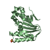
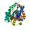
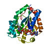
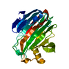
lytic cellulose monooxygenase (C4-dehydrogenating) / cellulose binding / cellulose catabolic process / monooxygenase activity / extracellular region / metal ion binding Similarity search - Function
Resolution: 1.6→69.64 Å / Cor.coef. Fo:Fc: 0.971 / Cor.coef. Fo:Fc free: 0.964 / SU B: 1.332 / SU ML: 0.047 / Cross valid method: THROUGHOUT / ESU R: 0.078 / ESU R Free: 0.077 / Stereochemistry target values: MAXIMUM LIKELIHOOD Details: HYDROGENS HAVE BEEN ADDED IN THE RIDING POSITIONS. U VALUES REFINED INDIVIDUALLY THE PROTEIN HAS BEEN TREATED WITH PAPAIN PRIOR TO CRYSTALLISATION. RESIDUES 175 TO 177 AND 209 TO 210 ARE ...Details: HYDROGENS HAVE BEEN ADDED IN THE RIDING POSITIONS. U VALUES REFINED INDIVIDUALLY THE PROTEIN HAS BEEN TREATED WITH PAPAIN PRIOR TO CRYSTALLISATION. RESIDUES 175 TO 177 AND 209 TO 210 ARE OMITTED DUE TO INSUFFICIENT ELECTRON DENSITY.
Rfactor
Num. reflection
% reflection
Selection details
Rfree
0.17808
1485
5.1 %
RANDOM
Rwork
0.15369
-
-
-
obs
0.15491
27351
99.99 %
-
Solvent computation
Ion probe radii: 0.8 Å / Shrinkage radii: 0.8 Å / VDW probe radii: 1.2 Å / Solvent model: MASK
 Movie
Movie Controller


 Open data
 Basic information
 Components
 Keywords
 Function and homology information
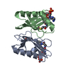
 NEUROSPORA CRASSA (fungus)
 X-RAY DIFFRACTION /
  Authors
 Citation
 Structure visualization
 Downloads & links
 Other downloads


 PDBj
PDBj


 Assembly

 Type: D-saccharide, alpha linking / Mass: 180.156 Da / Num. of mol.: 2


 Mass: 63.546 Da / Num. of mol.: 1 / Source method: obtained synthetically / Formula: Cu
 Mass: 96.063 Da / Num. of mol.: 3 / Source method: obtained synthetically / Formula: SO4
 Mass: 6.941 Da / Num. of mol.: 1 / Source method: obtained synthetically / Formula: Li
 Sample preparation
 / Beamline: ID23-2 / Wavelength: 0.8726
  Processing