 Movie
Movie Controller

[English] 日本語
 Yorodumi
- PDB-5fio: DARPins as a new tool for experimental phasing in protein crystal... -
+ Open data

- Basic information
| Entry | Database: PDB / ID: 5fio | |||||||||
|---|---|---|---|---|---|---|---|---|---|---|
| Title | DARPins as a new tool for experimental phasing in protein crystallography | |||||||||
 Components Components |
| |||||||||
 Keywords Keywords | METAL BINDING PROTEIN / DARPIN / ENGINEERED PROTEIN / EXPERIMENTAL PHASING | |||||||||
| Function / homology |  Function and homology information Function and homology informationdetection of maltose stimulus / maltose transport complex / carbohydrate transport / carbohydrate transmembrane transporter activity / maltose binding / maltose transport / maltodextrin transmembrane transport / ATP-binding cassette (ABC) transporter complex, substrate-binding subunit-containing / ATP-binding cassette (ABC) transporter complex / cell chemotaxis ...detection of maltose stimulus / maltose transport complex / carbohydrate transport / carbohydrate transmembrane transporter activity / maltose binding / maltose transport / maltodextrin transmembrane transport / ATP-binding cassette (ABC) transporter complex, substrate-binding subunit-containing / ATP-binding cassette (ABC) transporter complex / cell chemotaxis / outer membrane-bounded periplasmic space / periplasmic space / DNA damage response / membrane Similarity search - Function | |||||||||
| Biological species | SYNTHETIC CONSTRUCT (others) | |||||||||
| Method |  X-RAY DIFFRACTION / SYNCHROTRON / SAD / Resolution: 2.1 Å X-RAY DIFFRACTION / SYNCHROTRON / SAD / Resolution: 2.1 Å | |||||||||
 Authors Authors | Batyuk, A. / Honegger, A. / Andres, F. / Briand, C. / Gruetter, M. / Plueckthun, A. | |||||||||
 Citation Citation | Journal: To be Published Title: Darpins as a New Tool for Experimental Phasing in Protein Crystallography Authors: Batyuk, A. / Honegger, A. / Andres, F. / Briand, C. / Gruetter, M. / Plueckthun, A. | |||||||||
| History |
|
- Structure visualization
| Structure viewer | Molecule: MolmilJmol/JSmol |
|---|
- Downloads & links
-Download
| PDBx/mmCIF format | 5fio.cif.gz | 301.6 KB | Display | PDBx/mmCIF format |
|---|---|---|---|---|
| PDB format | pdb5fio.ent.gz | 250.2 KB | Display | PDB format |
| PDBx/mmJSON format | 5fio.json.gz | Tree view | PDBx/mmJSON format | |
| Others |  Other downloads Other downloads |
-Validation report
| Arichive directory | https://data.pdbj.org/pub/pdb/validation_reports/fi/5fioftp://data.pdbj.org/pub/pdb/validation_reports/fi/5fio | HTTPS FTP |
|---|
-Related structure data


-Links
 PDBj
PDBj


- Assembly
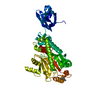
| Deposited unit | 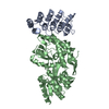
| ||||||||
|---|---|---|---|---|---|---|---|---|---|
| 1 |
| ||||||||
| Unit cell |
|
-Components
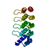
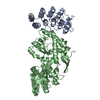
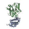
| #1: Protein | Mass: 18583.836 Da / Num. of mol.: 1 Source method: isolated from a genetically manipulated source Details: DESIGNED ANKYRIN REPEAT PROTEIN WITH THREE INTERNAL REPEAT AND C-TERMINAL CAPPING REPEAT TYPE MUT5 AND ENGINEERED BURIED MERCURY BINDING SITE CYS30-CYS65 WITH BOUND HG-ION Source: (gene. exp.) SYNTHETIC CONSTRUCT (others) / Plasmid: PQE30SS / Production host: |
|---|---|
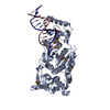
| #2: Protein | Mass: 43187.715 Da / Num. of mol.: 1 / Fragment: UNP RESIDUES 29-392 Source method: isolated from a genetically manipulated source Details: INVOLVED IN THE HIGH-AFFINITY MALTOSE MEMBRANE TRANSPORT SYSTEM MALEFGK. INITIAL RECEPTOR FOR THE ACTIVE TRANSPORT OF AND CHEMOTAXIS TOWARD MALTOOLIGOSACCHARIDES. Source: (gene. exp.) |
| #3: Chemical | ChemComp-HG /   Mass: 200.590 Da / Num. of mol.: 1 / Source method: obtained synthetically / Formula: Hg Mass: 200.590 Da / Num. of mol.: 1 / Source method: obtained synthetically / Formula: Hg |
| #4: Water | ChemComp-HOH /  Mass: 18.015 Da / Num. of mol.: 202 / Source method: isolated from a natural source / Formula: H2O Mass: 18.015 Da / Num. of mol.: 202 / Source method: isolated from a natural source / Formula: H2O |
-Experimental details
-Experiment
| Experiment | Method: X-RAY DIFFRACTION / Number of used crystals: 1 |
|---|
- Sample preparation
| Crystal | Density Matthews: 2.14 Å3/Da / Density % sol: 42.64 % / Description: NONE |
|---|---|
| Crystal grow | pH: 8 / Details: 30.55% W/V PEG 6000, 0.1M TRIS-CL, PH 8.0 |
-Data collection
| Diffraction | Mean temperature: 100 K |
|---|---|
| Diffraction source | Source: SYNCHROTRON / Site: SLS  / Beamline: X06SA / Wavelength: 1.00745 / Beamline: X06SA / Wavelength: 1.00745 |
| Detector | Type: DECTRIS PILATUS 6M / Detector: PIXEL / Date: Jun 7, 2011 / Details: RH COATED MERIDIONALLY FOCUSSING MIRROR |
| Radiation | Monochromator: FIXED-EXIT LN2 COOLED DOUBLE CRYSTAL MONOCHROMATOR Protocol: SINGLE WAVELENGTH / Monochromatic (M) / Laue (L): M / Scattering type: x-ray |
| Radiation wavelength | Wavelength: 1.00745 Å / Relative weight: 1 |
| Reflection | Resolution: 2.1→45.18 Å / Num. obs: 28809 / % possible obs: 98.9 % / Observed criterion σ(I): 2 / Redundancy: 2.9 % / Biso Wilson estimate: 32 Å2 / Rmerge(I) obs: 0.07 / Net I/σ(I): 12.92 |
| Reflection shell | Highest resolution: 2.1 Å / Redundancy: 2.9 % / Rmerge(I) obs: 0.55 / Mean I/σ(I) obs: 2.21 / % possible all: 98.2 |
- Processing
| Software |
| |||||||||||||||||||||||||||||||||||||||||||||||||||||||||||||||||||||||||||||||||||||||||||||||||||||||||||||||||||||||||||||||||||||||||||||||||||||||||||||||||||||||||||||||||||||||||||||||||||||||||||||||||||||||||||||||||
|---|---|---|---|---|---|---|---|---|---|---|---|---|---|---|---|---|---|---|---|---|---|---|---|---|---|---|---|---|---|---|---|---|---|---|---|---|---|---|---|---|---|---|---|---|---|---|---|---|---|---|---|---|---|---|---|---|---|---|---|---|---|---|---|---|---|---|---|---|---|---|---|---|---|---|---|---|---|---|---|---|---|---|---|---|---|---|---|---|---|---|---|---|---|---|---|---|---|---|---|---|---|---|---|---|---|---|---|---|---|---|---|---|---|---|---|---|---|---|---|---|---|---|---|---|---|---|---|---|---|---|---|---|---|---|---|---|---|---|---|---|---|---|---|---|---|---|---|---|---|---|---|---|---|---|---|---|---|---|---|---|---|---|---|---|---|---|---|---|---|---|---|---|---|---|---|---|---|---|---|---|---|---|---|---|---|---|---|---|---|---|---|---|---|---|---|---|---|---|---|---|---|---|---|---|---|---|---|---|---|---|---|---|---|---|---|---|---|---|---|---|---|---|---|---|---|---|
| Refinement | Method to determine structure: SAD Starting model: NONE Resolution: 2.1→45.178 Å / SU ML: 0.28 / σ(F): 0.86 / Phase error: 27.46 / Stereochemistry target values: MLHL
| |||||||||||||||||||||||||||||||||||||||||||||||||||||||||||||||||||||||||||||||||||||||||||||||||||||||||||||||||||||||||||||||||||||||||||||||||||||||||||||||||||||||||||||||||||||||||||||||||||||||||||||||||||||||||||||||||
| Solvent computation | Shrinkage radii: 1 Å / VDW probe radii: 1.2 Å / Solvent model: FLAT BULK SOLVENT MODEL | |||||||||||||||||||||||||||||||||||||||||||||||||||||||||||||||||||||||||||||||||||||||||||||||||||||||||||||||||||||||||||||||||||||||||||||||||||||||||||||||||||||||||||||||||||||||||||||||||||||||||||||||||||||||||||||||||
| Displacement parameters | Biso mean: 41.1 Å2 | |||||||||||||||||||||||||||||||||||||||||||||||||||||||||||||||||||||||||||||||||||||||||||||||||||||||||||||||||||||||||||||||||||||||||||||||||||||||||||||||||||||||||||||||||||||||||||||||||||||||||||||||||||||||||||||||||
| Refinement step | Cycle: LAST / Resolution: 2.1→45.178 Å
| |||||||||||||||||||||||||||||||||||||||||||||||||||||||||||||||||||||||||||||||||||||||||||||||||||||||||||||||||||||||||||||||||||||||||||||||||||||||||||||||||||||||||||||||||||||||||||||||||||||||||||||||||||||||||||||||||
| Refine LS restraints |
| |||||||||||||||||||||||||||||||||||||||||||||||||||||||||||||||||||||||||||||||||||||||||||||||||||||||||||||||||||||||||||||||||||||||||||||||||||||||||||||||||||||||||||||||||||||||||||||||||||||||||||||||||||||||||||||||||
| LS refinement shell |
| |||||||||||||||||||||||||||||||||||||||||||||||||||||||||||||||||||||||||||||||||||||||||||||||||||||||||||||||||||||||||||||||||||||||||||||||||||||||||||||||||||||||||||||||||||||||||||||||||||||||||||||||||||||||||||||||||
| Refinement TLS params. | Method: refined / Refine-ID: X-RAY DIFFRACTION
| |||||||||||||||||||||||||||||||||||||||||||||||||||||||||||||||||||||||||||||||||||||||||||||||||||||||||||||||||||||||||||||||||||||||||||||||||||||||||||||||||||||||||||||||||||||||||||||||||||||||||||||||||||||||||||||||||
| Refinement TLS group |
|
