 Movie
Movie Controller

[English] 日本語
 Yorodumi
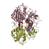
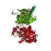
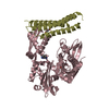
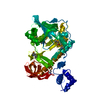
- PDB-5fgu: Structure of Sda1 nuclease apoprotein as an EGFP fixed-arm fusion -
+ Open data

- Basic information
| Entry | Database: PDB / ID: 5fgu | |||||||||
|---|---|---|---|---|---|---|---|---|---|---|
| Title | Structure of Sda1 nuclease apoprotein as an EGFP fixed-arm fusion | |||||||||
 Components Components | Green fluorescent protein,Extracellular streptodornase D | |||||||||
 Keywords Keywords | Metal binding / DNA binding protein / beta-beta-alpha metal finger nuclease / sequence nonspecific DNA binding / Metal binding protein | |||||||||
| Function / homology |  Function and homology information Function and homology informationsymbiont-mediated disruption of host neutrophil extracellular traps / bioluminescence / generation of precursor metabolites and energy / nucleic acid binding / hydrolase activity / metal ion binding Similarity search - Function | |||||||||
| Biological species |   Aequorea victoria (jellyfish) Aequorea victoria (jellyfish) Streptococcus pyogenes (bacteria) Streptococcus pyogenes (bacteria) | |||||||||
| Method |  X-RAY DIFFRACTION / SYNCHROTRON / MOLECULAR REPLACEMENT / Resolution: 1.896 Å X-RAY DIFFRACTION / SYNCHROTRON / MOLECULAR REPLACEMENT / Resolution: 1.896 Å | |||||||||
 Authors Authors | Moon, A.F. / Krahn, J.M. / Xun, L. / Cuneo, M.J. / Pedersen, L.C. | |||||||||
| Funding support |  United States, 1items United States, 1items
| |||||||||
 Citation Citation | Journal: Nucleic Acids Res. / Year: 2016 Title: Structural characterization of the virulence factor Sda1 nuclease from Streptococcus pyogenes. Authors: Moon, A.F. / Krahn, J.M. / Lu, X. / Cuneo, M.J. / Pedersen, L.C. | |||||||||
| History |
|
- Structure visualization
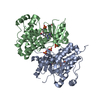
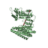
| Structure viewer | Molecule: MolmilJmol/JSmol |
|---|
- Downloads & links
-Download
| PDBx/mmCIF format | 5fgu.cif.gz | 125 KB | Display | PDBx/mmCIF format |
|---|---|---|---|---|
| PDB format | pdb5fgu.ent.gz | 93.3 KB | Display | PDB format |
| PDBx/mmJSON format | 5fgu.json.gz | Tree view | PDBx/mmJSON format | |
| Others |  Other downloads Other downloads |
-Validation report
| Arichive directory | https://data.pdbj.org/pub/pdb/validation_reports/fg/5fguftp://data.pdbj.org/pub/pdb/validation_reports/fg/5fgu | HTTPS FTP |
|---|
-Related structure data
| Related structure data |  5fgwC  4jrbS C: citing same article ( S: Starting model for refinement |
|---|---|
| Similar structure data |


-Links
 PDBj
PDBj

- Assembly
| Deposited unit | 
| ||||||||
|---|---|---|---|---|---|---|---|---|---|
| 1 |
| ||||||||
| 2 | 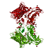
| ||||||||
| Unit cell |
| ||||||||
| Components on special symmetry positions |
| ||||||||
| Details | The target protein was shown by SANS to be monomeric in solution. |
-Components
| #1: Protein | Mass: 63627.629 Da / Num. of mol.: 1 Source method: isolated from a genetically manipulated source Source: (gene. exp.) Aequorea victoria (jellyfish), (gene. exp.) Streptococcus pyogenes (bacteria)Gene: GFP, sda1, sdaD2, HKU360_01468, SD90_06600 / Plasmid: pET32bEGFPX / Details (production host): EGFP fixed-arm fusion / Production host: | ||||||||
|---|---|---|---|---|---|---|---|---|---|
| #2: Chemical | ChemComp-SO4 /   Mass: 96.063 Da / Num. of mol.: 6 / Source method: obtained synthetically / Formula: SO4 Mass: 96.063 Da / Num. of mol.: 6 / Source method: obtained synthetically / Formula: SO4#3: Chemical | ChemComp-EDO /   Mass: 62.068 Da / Num. of mol.: 4 / Source method: obtained synthetically / Formula: C2H6O2 Mass: 62.068 Da / Num. of mol.: 4 / Source method: obtained synthetically / Formula: C2H6O2#4: Chemical | ChemComp-ACT /   Mass: 59.044 Da / Num. of mol.: 4 / Source method: obtained synthetically / Formula: C2H3O2 Mass: 59.044 Da / Num. of mol.: 4 / Source method: obtained synthetically / Formula: C2H3O2#5: Water | ChemComp-HOH / |  Mass: 18.015 Da / Num. of mol.: 327 / Source method: isolated from a natural source / Formula: H2O Mass: 18.015 Da / Num. of mol.: 327 / Source method: isolated from a natural source / Formula: H2OHas protein modification | Y | |
-Experimental details
-Experiment
| Experiment | Method: X-RAY DIFFRACTION |
|---|
- Sample preparation
| Crystal | Density Matthews: 2.7 Å3/Da / Density % sol: 54.5 % / Description: bullet-shaped |
|---|---|
| Crystal grow | Temperature: 277 K / Method: vapor diffusion, sitting drop / pH: 6 Details: Crystals were grown by mixing 0.25uL of protein (13.3mg/mL) with 0.25uL mother liquor (45mM Na cacodylate pH 6, 13.5mM magnesium sulfate, 1.53M ammonium sulfate), using sitting drop vapor diffusion |
-Data collection
| Diffraction | Mean temperature: 93.15 K |
|---|---|
| Diffraction source | Source: SYNCHROTRON / Site: APS / Beamline: 22-ID / Wavelength: 1 Å |
| Detector | Type: RAYONIX MX300-HS / Detector: CCD / Date: Feb 19, 2015 |
| Radiation | Protocol: SINGLE WAVELENGTH / Monochromatic (M) / Laue (L): M / Scattering type: x-ray |
| Radiation wavelength | Wavelength: 1 Å / Relative weight: 1 |
| Reflection | Resolution: 1.896→50 Å / Num. all: 54586 / Num. obs: 54586 / % possible obs: 100 % / Redundancy: 7.3 % / Rsym value: 0.092 / Net I/σ(I): 25.6 |
| Reflection shell | Resolution: 1.896→1.93 Å / Redundancy: 6.4 % / Rmerge(I) obs: 0.61 / Mean I/σ(I) obs: 3.54 / % possible all: 99.6 |
- Processing
| Software |
| |||||||||||||||||||||||||||||||||||||||||||||||||||||||||||||||||||||||||||||||||||||||||||||||||||||||||||||||||||||||||||||||||||||||||||||||||||
|---|---|---|---|---|---|---|---|---|---|---|---|---|---|---|---|---|---|---|---|---|---|---|---|---|---|---|---|---|---|---|---|---|---|---|---|---|---|---|---|---|---|---|---|---|---|---|---|---|---|---|---|---|---|---|---|---|---|---|---|---|---|---|---|---|---|---|---|---|---|---|---|---|---|---|---|---|---|---|---|---|---|---|---|---|---|---|---|---|---|---|---|---|---|---|---|---|---|---|---|---|---|---|---|---|---|---|---|---|---|---|---|---|---|---|---|---|---|---|---|---|---|---|---|---|---|---|---|---|---|---|---|---|---|---|---|---|---|---|---|---|---|---|---|---|---|---|---|---|
| Refinement | Method to determine structure: MOLECULAR REPLACEMENT Starting model: EGFP component of 4JRB Resolution: 1.896→38.276 Å / SU ML: 0.17 / Cross valid method: FREE R-VALUE / σ(F): 1.36 / Phase error: 21.93 / Stereochemistry target values: ML Details: The authors state that the vast majority of the outliers in the structure come from regions that are not well ordered--especially residues from the C-terminal domain (residues 1336-1385). ...Details: The authors state that the vast majority of the outliers in the structure come from regions that are not well ordered--especially residues from the C-terminal domain (residues 1336-1385). This domain is largely disordered, with the exception of a few small snippets of secondary structure. These regions are not well ordered and the density is very weak, but enough to assign sequence. Occupancy refinement of these residues suggests they are present at full occupancy.
| |||||||||||||||||||||||||||||||||||||||||||||||||||||||||||||||||||||||||||||||||||||||||||||||||||||||||||||||||||||||||||||||||||||||||||||||||||
| Solvent computation | Shrinkage radii: 0.9 Å / VDW probe radii: 1.11 Å / Solvent model: FLAT BULK SOLVENT MODEL | |||||||||||||||||||||||||||||||||||||||||||||||||||||||||||||||||||||||||||||||||||||||||||||||||||||||||||||||||||||||||||||||||||||||||||||||||||
| Refinement step | Cycle: LAST / Resolution: 1.896→38.276 Å
| |||||||||||||||||||||||||||||||||||||||||||||||||||||||||||||||||||||||||||||||||||||||||||||||||||||||||||||||||||||||||||||||||||||||||||||||||||
| Refine LS restraints |
| |||||||||||||||||||||||||||||||||||||||||||||||||||||||||||||||||||||||||||||||||||||||||||||||||||||||||||||||||||||||||||||||||||||||||||||||||||
| LS refinement shell |
|
