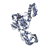
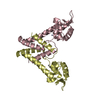
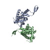
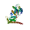
[English] 日本語
Yorodumi
- PDB-5fd6: zinc-bound manganese uptake regulator -

+
Open data


ID or keywords:

Loading...

-
Basic information

Entry
Database: PDB / ID: 5fd6
Titlezinc-bound manganese uptake regulator
ComponentsFerric uptake regulation protein
KeywordsTRANSCRIPTION / manganese uptake regulator / fur / mur / ferric uptake regulator
Function / homology
Function and homology information


DNA-binding transcription factor activity / DNA binding / metal ion binding / cytoplasm
Similarity search - Function
Ferric-uptake regulator, C-terminal dimerisarion domain / Ferric-uptake regulator, C-terminal domain / Ferric-uptake regulator / Ferric uptake regulator family / Dna Ligase; domain 1 / Winged helix-like DNA-binding domain superfamily/Winged helix DNA-binding domain / Arc Repressor Mutant, subunit A / Winged helix DNA-binding domain superfamily / Winged helix-like DNA-binding domain superfamily / 2-Layer Sandwich ...Ferric-uptake regulator, C-terminal dimerisarion domain / Ferric-uptake regulator, C-terminal domain / Ferric-uptake regulator / Ferric uptake regulator family / Dna Ligase; domain 1 / Winged helix-like DNA-binding domain superfamily/Winged helix DNA-binding domain / Arc Repressor Mutant, subunit A / Winged helix DNA-binding domain superfamily / Winged helix-like DNA-binding domain superfamily / 2-Layer Sandwich / Orthogonal Bundle / Mainly Alpha / Alpha Beta
Similarity search - Domain/homology
Ferric uptake regulation protein
Similarity search - Component
Biological speciesRhizobium leguminosarum bv. viciae (bacteria)
MethodX-RAY DIFFRACTION / SYNCHROTRON / MOLECULAR REPLACEMENT / Resolution: 2.48 Å
AuthorsBellini, D. / Hemmings, A.M. / Walsh, M.A.
Funding support United Kingdom, 1items
OrganizationGrant numberCountry
United Kingdom
CitationJournal: To Be Published
Title: structure of a zinc-bound manganese uptake regulator, mur
Authors: Bellini, D. / Lebedev, A. / Keegan, R. / Todd, J. / Hemmings, A.M. / Johnston, A.W. / Walsh, M.A.
History
DepositionDec 15, 2015Deposition site: RCSB / Processing site: PDBE
Revision 1.0Dec 28, 2016Provider: repository / Type: Initial release
Revision 1.1May 8, 2024Group: Data collection / Database references / Category: chem_comp_atom / chem_comp_bond / database_2
Item: _database_2.pdbx_DOI / _database_2.pdbx_database_accession

-
Structure visualization

Structure viewerMolecule:
MolmilJmol/JSmol

Downloads & links

-
Assembly

Deposited unit
A: Ferric uptake regulation protein
B: Ferric uptake regulation protein
C: Ferric uptake regulation protein
D: Ferric uptake regulation protein
hetero molecules


Theoretical massNumber of molelcules
Total (without water)67,94318
Polymers66,8484
Non-polymers1,09614
Water19811
1
A: Ferric uptake regulation protein
B: Ferric uptake regulation protein
hetero molecules


Theoretical massNumber of molelcules
Total (without water)34,07010
Polymers33,4242
Non-polymers6468
Water181
TypeNameSymmetry operationNumber
identity operation1_555x,y,z1
Buried area3760 Å2
ΔGint-77 kcal/mol
Surface area15370 Å2
MethodPISA
2
C: Ferric uptake regulation protein
D: Ferric uptake regulation protein
hetero molecules


Theoretical massNumber of molelcules
Total (without water)33,8748
Polymers33,4242
Non-polymers4506
Water362
TypeNameSymmetry operationNumber
identity operation1_555x,y,z1
Buried area3050 Å2
ΔGint-33 kcal/mol
Surface area15310 Å2
MethodPISA
Unit cell
Length a, b, c (Å)94.950, 94.950, 63.020
Angle α, β, γ (deg.)90.00, 90.00, 90.00
Int Tables number78
Space group name H-MP43

-
Components

#1: Protein
Ferric uptake regulation protein / Ferric uptake regulator


Mass: 16711.914 Da / Num. of mol.: 4 / Fragment: UNP residues 2-142
Source method: isolated from a genetically manipulated source
Source: (gene. exp.) Rhizobium leguminosarum bv. viciae (bacteria)
Gene: fur / Production host: Escherichia coli (E. coli) / References: UniProt: O07315
#2: Chemical
ChemComp-ZN / ZINC ION


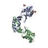
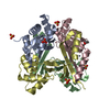
Mass: 65.409 Da / Num. of mol.: 8 / Source method: obtained synthetically / Formula: Zn
#3: Chemical
ChemComp-SO4 / SULFATE ION


Mass: 96.063 Da / Num. of mol.: 5 / Source method: obtained synthetically / Formula: SO4
#4: Chemical ChemComp-GOL / GLYCEROL / GLYCERIN / PROPANE-1,2,3-TRIOL


Mass: 92.094 Da / Num. of mol.: 1 / Source method: obtained synthetically / Formula: C3H8O3
#5: Water ChemComp-HOH / water


Mass: 18.015 Da / Num. of mol.: 11 / Source method: isolated from a natural source / Formula: H2O

-
Experimental details

-
Experiment

ExperimentMethod: X-RAY DIFFRACTION / Number of used crystals: 1

-
Sample preparation

CrystalDensity Matthews: 2.19 Å3/Da / Density % sol: 43.86 %
Crystal growTemperature: 277 K / Method: vapor diffusion, hanging drop
Details: 0.1 M Trizma, pH 8.0, 1.5 M ammonium sulfate. The protein sample contained a 10:1 molar excess of Zn(II)

-
Data collection

DiffractionMean temperature: 100 K
Diffraction sourceSource: SYNCHROTRON / Site: SRS / Beamline: PX9.6 / Wavelength: 0.933 Å
DetectorType: ADSC QUANTUM 315r / Detector: CCD / Date: Sep 15, 2003
RadiationProtocol: SINGLE WAVELENGTH / Monochromatic (M) / Laue (L): M / Scattering type: x-ray
Radiation wavelengthWavelength: 0.933 Å / Relative weight: 1
Reflection twin
Crystal-IDIDOperatorDomain-IDFraction
11H, K, L10.68
11-H, K, -L20.32
ReflectionResolution: 2.5→94.9 Å / Num. obs: 19841 / % possible obs: 99.7 % / Redundancy: 2.1 % / Rmerge(I) obs: 0.092 / Net I/σ(I): 9.1
Reflection shellResolution: 2.5→2.6 Å / Rmerge(I) obs: 0.26 / Mean I/σ(I) obs: 4.2 / % possible all: 99.4

-
Processing

Software
NameVersionClassification
REFMAC5.8.0135refinement
HKL-2000data reduction
SCALEPACKdata scaling
MOLREPphasing
RefinementMethod to determine structure: MOLECULAR REPLACEMENT / Resolution: 2.48→42.46 Å / Cor.coef. Fo:Fc: 0.951 / Cor.coef. Fo:Fc free: 0.938 / SU B: 8.583 / SU ML: 0.202 / Cross valid method: THROUGHOUT / ESU R: 0.188 / ESU R Free: 0.058 / Stereochemistry target values: MAXIMUM LIKELIHOOD / Details: HYDROGENS HAVE BEEN ADDED IN THE RIDING POSITIONS
RfactorNum. reflection% reflectionSelection details
Rfree0.2245 993 5 %RANDOM
Rwork0.18724 ---
obs0.18891 19064 99.67 %-
Solvent computationIon probe radii: 0.8 Å / Shrinkage radii: 0.8 Å / VDW probe radii: 1.2 Å / Solvent model: MASK
Displacement parametersBiso mean: 68.556 Å2
Baniso -1Baniso -2Baniso -3
1-7.19 Å20 Å20 Å2
2--7.19 Å20 Å2
3----14.38 Å2
Refinement stepCycle: LAST / Resolution: 2.48→42.46 Å
ProteinNucleic acidLigandSolventTotal
Num. atoms4400 0 39 11 4450
Refine LS restraints
Refine-IDTypeDev idealDev ideal targetNumber
X-RAY DIFFRACTIONr_bond_refined_d0.0090.0194497
X-RAY DIFFRACTIONr_bond_other_d0.0040.024350
X-RAY DIFFRACTIONr_angle_refined_deg1.4291.9716053
X-RAY DIFFRACTIONr_angle_other_deg0.93239980
X-RAY DIFFRACTIONr_dihedral_angle_1_deg7.055528
X-RAY DIFFRACTIONr_dihedral_angle_2_deg27.25622.195246
X-RAY DIFFRACTIONr_dihedral_angle_3_deg15.9915846
X-RAY DIFFRACTIONr_dihedral_angle_4_deg14.0281566
X-RAY DIFFRACTIONr_chiral_restr0.0730.2674
X-RAY DIFFRACTIONr_gen_planes_refined0.0050.024946
X-RAY DIFFRACTIONr_gen_planes_other0.0020.021048
X-RAY DIFFRACTIONr_nbd_refined
X-RAY DIFFRACTIONr_nbd_other
X-RAY DIFFRACTIONr_nbtor_refined
X-RAY DIFFRACTIONr_nbtor_other
X-RAY DIFFRACTIONr_xyhbond_nbd_refined
X-RAY DIFFRACTIONr_xyhbond_nbd_other
X-RAY DIFFRACTIONr_metal_ion_refined
X-RAY DIFFRACTIONr_metal_ion_other
X-RAY DIFFRACTIONr_symmetry_vdw_refined
X-RAY DIFFRACTIONr_symmetry_vdw_other
X-RAY DIFFRACTIONr_symmetry_hbond_refined
X-RAY DIFFRACTIONr_symmetry_hbond_other
X-RAY DIFFRACTIONr_symmetry_metal_ion_refined
X-RAY DIFFRACTIONr_symmetry_metal_ion_other
X-RAY DIFFRACTIONr_mcbond_it3.9946.5772124
X-RAY DIFFRACTIONr_mcbond_other3.9896.5772123
X-RAY DIFFRACTIONr_mcangle_it6.1199.8582648
X-RAY DIFFRACTIONr_mcangle_other6.1199.8582649
X-RAY DIFFRACTIONr_scbond_it3.9837.0432373
X-RAY DIFFRACTIONr_scbond_other3.9837.0432373
X-RAY DIFFRACTIONr_scangle_it
X-RAY DIFFRACTIONr_scangle_other6.3110.3783406
X-RAY DIFFRACTIONr_long_range_B_refined9.71550.5814866
X-RAY DIFFRACTIONr_long_range_B_other9.71550.5824866
X-RAY DIFFRACTIONr_rigid_bond_restr
X-RAY DIFFRACTIONr_sphericity_free
X-RAY DIFFRACTIONr_sphericity_bonded
LS refinement shellResolution: 2.478→2.543 Å / Total num. of bins used: 20
RfactorNum. reflection% reflection
Rfree0.295 106 -
Rwork0.217 1354 -
obs--98.58 %

+
About Yorodumi

-
News

-
Feb 9, 2022. New format data for meta-information of EMDB entries

New format data for meta-information of EMDB entries

  • Version 3 of the EMDB header file is now the official format.
  • The previous official version 1.9 will be removed from the archive.

Related info.:EMDB header

External links:wwPDB to switch to version 3 of the EMDB data model

-
Aug 12, 2020. Covid-19 info

Covid-19 info

URL: https://pdbjlvh1.pdbj.org/emnavi/covid19.php

New page: Covid-19 featured information page in EM Navigator.

Related info.:Covid-19 info / Mar 5, 2020. Novel coronavirus structure data

+
Mar 5, 2020. Novel coronavirus structure data

Novel coronavirus structure data

Related info.:Yorodumi Speices / Aug 12, 2020. Covid-19 info

External links:COVID-19 featured content - PDBj / Molecule of the Month (242):Coronavirus Proteases

+
Jan 31, 2019. EMDB accession codes are about to change! (news from PDBe EMDB page)

EMDB accession codes are about to change! (news from PDBe EMDB page)

  • The allocation of 4 digits for EMDB accession codes will soon come to an end. Whilst these codes will remain in use, new EMDB accession codes will include an additional digit and will expand incrementally as the available range of codes is exhausted. The current 4-digit format prefixed with “EMD-” (i.e. EMD-XXXX) will advance to a 5-digit format (i.e. EMD-XXXXX), and so on. It is currently estimated that the 4-digit codes will be depleted around Spring 2019, at which point the 5-digit format will come into force.
  • The EM Navigator/Yorodumi systems omit the EMD- prefix.

Related info.:Q: What is EMD? / ID/Accession-code notation in Yorodumi/EM Navigator

External links:EMDB Accession Codes are Changing Soon! / Contact to PDBj

+
Jul 12, 2017. Major update of PDB

Major update of PDB

  • wwPDB released updated PDB data conforming to the new PDBx/mmCIF dictionary.
  • This is a major update changing the version number from 4 to 5, and with Remediation, in which all the entries are updated.
  • In this update, many items about electron microscopy experimental information are reorganized (e.g. em_software).
  • Now, EM Navigator and Yorodumi are based on the updated data.

External links:wwPDB Remediation / Enriched Model Files Conforming to OneDep Data Standards Now Available in the PDB FTP Archive

-
Yorodumi

Thousand views of thousand structures

  • Yorodumi is a browser for structure data from EMDB, PDB, SASBDB, etc.
  • This page is also the successor to EM Navigator detail page, and also detail information page/front-end page for Omokage search.
  • The word "yorodu" (or yorozu) is an old Japanese word meaning "ten thousand". "mi" (miru) is to see.

Related info.:EMDB / PDB / SASBDB / Comparison of 3 databanks / Yorodumi Search / Aug 31, 2016. New EM Navigator & Yorodumi / Yorodumi Papers / Jmol/JSmol / Function and homology information / Changes in new EM Navigator and Yorodumi

Read more