 Movie
Movie Controller

[English] 日本語
 Yorodumi
- PDB-5fa1: The structure of the beta-3-deoxy-D-manno-oct-2-ulosonic acid tra... -
+ Open data

- Basic information
| Entry | Database: PDB / ID: 5fa1 | ||||||
|---|---|---|---|---|---|---|---|
| Title | The structure of the beta-3-deoxy-D-manno-oct-2-ulosonic acid transferase domain of WbbB | ||||||
 Components Components | Putative N-acetyl glucosaminyl transferase | ||||||
 Keywords Keywords | TRANSFERASE / LPS biosynthesis / glycosyltransferase | ||||||
| Function / homology | : / Glycosyltransferase 99 family N-terminal domain / Capsule polysaccharide biosynthesis / Capsule polysaccharide biosynthesis protein / polysaccharide transport / polysaccharide biosynthetic process / transferase activity / CYTIDINE-5'-MONOPHOSPHATE / Putative N-acetyl glucosaminyl transferase Function and homology information Function and homology information | ||||||
| Biological species |  Raoultella terrigena (bacteria) Raoultella terrigena (bacteria) | ||||||
| Method |  X-RAY DIFFRACTION / SYNCHROTRON / MOLECULAR REPLACEMENT / Resolution: 2.1 Å X-RAY DIFFRACTION / SYNCHROTRON / MOLECULAR REPLACEMENT / Resolution: 2.1 Å | ||||||
 Authors Authors | Mallette, E. / Ovchinnikova, O.G. / Whitfield, C. / Kimber, M.S. | ||||||
| Funding support |  Canada, 1items Canada, 1items
| ||||||
 Citation Citation | Journal: Proc. Natl. Acad. Sci. U.S.A. / Year: 2016 Title: Bacterial beta-Kdo glycosyltransferases represent a new glycosyltransferase family (GT99). Authors: Ovchinnikova, O.G. / Mallette, E. / Koizumi, A. / Lowary, T.L. / Kimber, M.S. / Whitfield, C. | ||||||
| History |
|
- Structure visualization
| Structure viewer | Molecule: MolmilJmol/JSmol |
|---|
- Downloads & links
-Download
| PDBx/mmCIF format | 5fa1.cif.gz | 339.1 KB | Display | PDBx/mmCIF format |
|---|---|---|---|---|
| PDB format | pdb5fa1.ent.gz | 274.4 KB | Display | PDB format |
| PDBx/mmJSON format | 5fa1.json.gz | Tree view | PDBx/mmJSON format | |
| Others |  Other downloads Other downloads |
-Validation report
| Arichive directory | https://data.pdbj.org/pub/pdb/validation_reports/fa/5fa1ftp://data.pdbj.org/pub/pdb/validation_reports/fa/5fa1 | HTTPS FTP |
|---|
-Related structure data
| Related structure data |  5fa0SC S: Starting model for refinement C: citing same article ( |
|---|---|
| Similar structure data |


-Links
 PDBj
PDBj- Assembly
| Deposited unit | 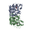
| |||||||||
|---|---|---|---|---|---|---|---|---|---|---|
| 1 |
| |||||||||
| Unit cell |
| |||||||||
| Components on special symmetry positions |
|
-Components
| #1: Protein | Mass: 46096.770 Da / Num. of mol.: 2 Source method: isolated from a genetically manipulated source Source: (gene. exp.) Raoultella terrigena (bacteria) / Gene: wbbB / Plasmid: pET28a(+) / Production host: #2: Chemical |   Mass: 323.197 Da / Num. of mol.: 2 / Source method: obtained synthetically / Formula: C9H14N3O8P Mass: 323.197 Da / Num. of mol.: 2 / Source method: obtained synthetically / Formula: C9H14N3O8P#3: Water | ChemComp-HOH / |  Mass: 18.015 Da / Num. of mol.: 837 / Source method: isolated from a natural source / Formula: H2O Mass: 18.015 Da / Num. of mol.: 837 / Source method: isolated from a natural source / Formula: H2O |
|---|
-Experimental details
-Experiment
| Experiment | Method: X-RAY DIFFRACTION |
|---|
- Sample preparation
| Crystal | Density Matthews: 2.4 Å3/Da / Density % sol: 48.9 % |
|---|---|
| Crystal grow | Temperature: 293 K / Method: vapor diffusion, sitting drop / pH: 5.5 Details: 10 mg/ml WbbB, 0.2 M NaCl, 0.1M Bis-Tris, 25% PEG 3350, 1 mM TCEP, 1mM CMP |
-Data collection
| Diffraction | Mean temperature: 100 K |
|---|---|
| Diffraction source | Source: SYNCHROTRON / Site: CLSI / Beamline: 08ID-1 / Wavelength: 0.97949 Å |
| Detector | Type: RAYONIX MX-300 / Detector: CCD / Date: Jul 10, 2015 |
| Radiation | Monochromator: ACCEL/BRUKER double crystal monochromator / Protocol: SINGLE WAVELENGTH / Monochromatic (M) / Laue (L): M / Scattering type: x-ray |
| Radiation wavelength | Wavelength: 0.97949 Å / Relative weight: 1 |
| Reflection | Resolution: 2.1→50 Å / Num. obs: 52154 / % possible obs: 100 % / Redundancy: 7.2 % / Rsym value: 0.094 / Net I/σ(I): 13.3 |
| Reflection shell | Resolution: 2.1→2.15 Å / Redundancy: 7.5 % / Rmerge(I) obs: 0.64 / Mean I/σ(I) obs: 2.85 / % possible all: 100 |
- Processing
| Software |
| |||||||||||||||||||||||||||||||||||||||||||||||||||||||||||||||||||||||||||||||||||||||||||||||||||||||||||||||||||||||||||||||||||||||||||||||||||||||||||||||||||||||||||||||||||||||||||||||||||||||||||||||||||||||||||||||||
|---|---|---|---|---|---|---|---|---|---|---|---|---|---|---|---|---|---|---|---|---|---|---|---|---|---|---|---|---|---|---|---|---|---|---|---|---|---|---|---|---|---|---|---|---|---|---|---|---|---|---|---|---|---|---|---|---|---|---|---|---|---|---|---|---|---|---|---|---|---|---|---|---|---|---|---|---|---|---|---|---|---|---|---|---|---|---|---|---|---|---|---|---|---|---|---|---|---|---|---|---|---|---|---|---|---|---|---|---|---|---|---|---|---|---|---|---|---|---|---|---|---|---|---|---|---|---|---|---|---|---|---|---|---|---|---|---|---|---|---|---|---|---|---|---|---|---|---|---|---|---|---|---|---|---|---|---|---|---|---|---|---|---|---|---|---|---|---|---|---|---|---|---|---|---|---|---|---|---|---|---|---|---|---|---|---|---|---|---|---|---|---|---|---|---|---|---|---|---|---|---|---|---|---|---|---|---|---|---|---|---|---|---|---|---|---|---|---|---|---|---|---|---|---|---|---|---|
| Refinement | Method to determine structure: MOLECULAR REPLACEMENT Starting model: 5FA0 Resolution: 2.1→48.046 Å / SU ML: 0.19 / Cross valid method: THROUGHOUT / σ(F): 1.36 / Phase error: 18.78 / Stereochemistry target values: ML
| |||||||||||||||||||||||||||||||||||||||||||||||||||||||||||||||||||||||||||||||||||||||||||||||||||||||||||||||||||||||||||||||||||||||||||||||||||||||||||||||||||||||||||||||||||||||||||||||||||||||||||||||||||||||||||||||||
| Solvent computation | Shrinkage radii: 0.9 Å / VDW probe radii: 1.11 Å / Solvent model: FLAT BULK SOLVENT MODEL | |||||||||||||||||||||||||||||||||||||||||||||||||||||||||||||||||||||||||||||||||||||||||||||||||||||||||||||||||||||||||||||||||||||||||||||||||||||||||||||||||||||||||||||||||||||||||||||||||||||||||||||||||||||||||||||||||
| Refinement step | Cycle: LAST / Resolution: 2.1→48.046 Å
| |||||||||||||||||||||||||||||||||||||||||||||||||||||||||||||||||||||||||||||||||||||||||||||||||||||||||||||||||||||||||||||||||||||||||||||||||||||||||||||||||||||||||||||||||||||||||||||||||||||||||||||||||||||||||||||||||
| Refine LS restraints |
| |||||||||||||||||||||||||||||||||||||||||||||||||||||||||||||||||||||||||||||||||||||||||||||||||||||||||||||||||||||||||||||||||||||||||||||||||||||||||||||||||||||||||||||||||||||||||||||||||||||||||||||||||||||||||||||||||
| LS refinement shell |
| |||||||||||||||||||||||||||||||||||||||||||||||||||||||||||||||||||||||||||||||||||||||||||||||||||||||||||||||||||||||||||||||||||||||||||||||||||||||||||||||||||||||||||||||||||||||||||||||||||||||||||||||||||||||||||||||||
| Refinement TLS params. | Method: refined / Refine-ID: X-RAY DIFFRACTION
| |||||||||||||||||||||||||||||||||||||||||||||||||||||||||||||||||||||||||||||||||||||||||||||||||||||||||||||||||||||||||||||||||||||||||||||||||||||||||||||||||||||||||||||||||||||||||||||||||||||||||||||||||||||||||||||||||
| Refinement TLS group |
|
