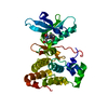
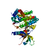
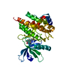
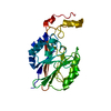
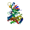
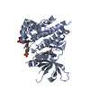
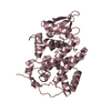
Entry Database : PDB / ID : 5f20Title Structure of TYK2 with inhibitor 4: 3-azanyl-5-(2-methylphenyl)-7-(1-methylpyrazol-3-yl)-1~{H}-pyrazolo[4,3-c]pyridin-4-one Non-receptor tyrosine-protein kinase TYK2 Keywords / / Function / homology Function Domain/homology Component
/ / / / / / / / / / / / / / / / / / / / / / / / / / / / / / / / / / / / / / / / / / / / / / / / / / / / / / / / / / / / / / / / / / / / / / / / / / / / / / / / / / / / / / / / / / / / / / / / / / / / / / / / / / / / / / / / Biological species Homo sapiens (human)Method / / / / Resolution : 2.91 Å Authors Skene, R.J. Journal : J.Med.Chem. / Year : 2016Title : Structure-Based Design and Synthesis of 3-Amino-1,5-dihydro-4H-pyrazolopyridin-4-one Derivatives as Tyrosine Kinase 2 Inhibitors.Authors: Yogo, T. / Nagamiya, H. / Seto, M. / Sasaki, S. / Shih-Chung, H. / Ohba, Y. / Tokunaga, N. / Lee, G.N. / Rhim, C.Y. / Yoon, C.H. / Cho, S.Y. / Skene, R. / Yamamoto, S. / Satou, Y. / Kuno, M. ... Authors : Yogo, T. / Nagamiya, H. / Seto, M. / Sasaki, S. / Shih-Chung, H. / Ohba, Y. / Tokunaga, N. / Lee, G.N. / Rhim, C.Y. / Yoon, C.H. / Cho, S.Y. / Skene, R. / Yamamoto, S. / Satou, Y. / Kuno, M. / Miyazaki, T. / Nakagawa, H. / Okabe, A. / Marui, S. / Aso, K. / Yoshida, M. History Deposition Dec 1, 2015 Deposition site / Processing site Revision 1.0 Jan 13, 2016 Provider / Type Revision 1.1 Jan 20, 2016 Group Revision 1.2 Feb 10, 2016 Group Revision 1.3 Mar 6, 2024 Group / Database references / Derived calculationsCategory chem_comp_atom / chem_comp_bond ... chem_comp_atom / chem_comp_bond / citation / database_2 / pdbx_struct_oper_list Item _citation.journal_id_CSD / _database_2.pdbx_DOI ... _citation.journal_id_CSD / _database_2.pdbx_DOI / _database_2.pdbx_database_accession / _pdbx_struct_oper_list.symmetry_operation
Show all Show less
 Movie
Movie Controller

 Yorodumi
 Open data
 Basic information
 Components
 Keywords
 Function and homology information
 Homo sapiens (human)
 X-RAY DIFFRACTION /
  Authors
 Citation
 Structure visualization
 Downloads & links
 Other downloads
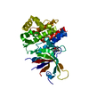


 PDBj
PDBj


 Assembly
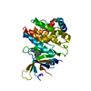
 unidentified baculovirus
 Mass: 320.349 Da / Num. of mol.: 1 / Source method: obtained synthetically / Formula: C17H16N6O
 Mass: 18.015 Da / Num. of mol.: 31 / Source method: isolated from a natural source / Formula: H2O
 Sample preparation
 / Beamline: 5.0.3 / Wavelength: 0.98 Å
 Processing