 Movie
Movie Controller

[English] 日本語
 Yorodumi
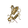
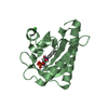
- PDB-5exh: Crystal structure of mTET3-CXXC domain in complex with 5-carboxyl... -
+ Open data

- Basic information
| Entry | Database: PDB / ID: 5exh | ||||||
|---|---|---|---|---|---|---|---|
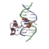
| Title | Crystal structure of mTET3-CXXC domain in complex with 5-carboxylcytosine DNA at 1.3 Angstroms resolution. | ||||||
 Components Components |
| ||||||
 Keywords Keywords | OXIDOREDUCTASE/DNA / mouse Tet3 / complex / 5-carboxylcytosine / reader / OXIDOREDUCTASE-DNA complex | ||||||
| Function / homology |  Function and homology information Function and homology informationepigenetic programing of male pronucleus / methylcytosine dioxygenase / DNA 5-methylcytosine dioxygenase activity / chromosomal 5-methylcytosine DNA demethylation, oxidation pathway / positive regulation of gene expression via chromosomal CpG island demethylation / protein O-linked glycosylation / male pronucleus / female pronucleus / methyl-CpG binding / chromosome ...epigenetic programing of male pronucleus / methylcytosine dioxygenase / DNA 5-methylcytosine dioxygenase activity / chromosomal 5-methylcytosine DNA demethylation, oxidation pathway / positive regulation of gene expression via chromosomal CpG island demethylation / protein O-linked glycosylation / male pronucleus / female pronucleus / methyl-CpG binding / chromosome / RNA polymerase II cis-regulatory region sequence-specific DNA binding / positive regulation of transcription by RNA polymerase II / nucleoplasm / zinc ion binding / nucleus / cytoplasm Similarity search - Function | ||||||
| Biological species |  synthetic construct (others) | ||||||
| Method |  X-RAY DIFFRACTION / SYNCHROTRON / MOLECULAR REPLACEMENT / Resolution: 1.3 Å X-RAY DIFFRACTION / SYNCHROTRON / MOLECULAR REPLACEMENT / Resolution: 1.3 Å | ||||||
 Authors Authors | Song, J. | ||||||
 Citation Citation | Journal: Cell Rep / Year: 2016 Title: Tet3 Reads 5-Carboxylcytosine through Its CXXC Domain and Is a Potential Guardian against Neurodegeneration. Authors: Jin, S.G. / Zhang, Z.M. / Dunwell, T.L. / Harter, M.R. / Wu, X. / Johnson, J. / Li, Z. / Liu, J. / Szabo, P.E. / Lu, Q. / Xu, G.L. / Song, J. / Pfeifer, G.P. | ||||||
| History |
|
- Structure visualization
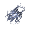
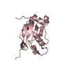
| Structure viewer | Molecule: MolmilJmol/JSmol |
|---|
- Downloads & links
-Download
| PDBx/mmCIF format | 5exh.cif.gz | 45.1 KB | Display | PDBx/mmCIF format |
|---|---|---|---|---|
| PDB format | pdb5exh.ent.gz | 27.5 KB | Display | PDB format |
| PDBx/mmJSON format | 5exh.json.gz | Tree view | PDBx/mmJSON format | |
| Others |  Other downloads Other downloads |
-Validation report
| Arichive directory | https://data.pdbj.org/pub/pdb/validation_reports/ex/5exhftp://data.pdbj.org/pub/pdb/validation_reports/ex/5exh | HTTPS FTP |
|---|
-Related structure data
| Related structure data |  4hp1S S: Starting model for refinement |
|---|---|
| Similar structure data |
-Links
 PDBj
PDBj


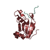
- Assembly
| Deposited unit | 
| ||||||||
|---|---|---|---|---|---|---|---|---|---|
| 1 |
| ||||||||
| Unit cell |
|
-Components
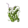
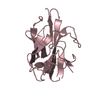
| #1: DNA chain | Mass: 3706.414 Da / Num. of mol.: 2 / Source method: obtained synthetically / Source: (synth.) synthetic construct (others) #2: Protein/peptide | | Mass: 5501.648 Da / Num. of mol.: 1 / Fragment: CXXC domain (UNP residues 51-96) Source method: isolated from a genetically manipulated source Source: (gene. exp.)  References: UniProt: L0HN04, UniProt: Q8BG87*PLUS, Oxidoreductases; Acting on paired donors, with incorporation or reduction of molecular oxygen; With 2-oxoglutarate as one donor, and incorporation ...References: UniProt: L0HN04, UniProt: Q8BG87*PLUS, Oxidoreductases; Acting on paired donors, with incorporation or reduction of molecular oxygen; With 2-oxoglutarate as one donor, and incorporation of one atom of oxygen into each donor #3: Chemical |   Mass: 65.409 Da / Num. of mol.: 2 / Source method: obtained synthetically / Formula: Zn Mass: 65.409 Da / Num. of mol.: 2 / Source method: obtained synthetically / Formula: Zn#4: Water | ChemComp-HOH / |  Mass: 18.015 Da / Num. of mol.: 230 / Source method: isolated from a natural source / Formula: H2O Mass: 18.015 Da / Num. of mol.: 230 / Source method: isolated from a natural source / Formula: H2O |
|---|
-Experimental details
-Experiment
| Experiment | Method: X-RAY DIFFRACTION / Number of used crystals: 1 |
|---|
- Sample preparation
| Crystal | Density Matthews: 1.8 Å3/Da / Density % sol: 31.69 % |
|---|---|
| Crystal grow | Temperature: 293 K / Method: vapor diffusion, hanging drop / pH: 7.5 Details: 0.1 M Hepes, pH 7.5, 50 mM, calcium chloride, 41% PEG200 |
-Data collection
| Diffraction | Mean temperature: 100 K |
|---|---|
| Diffraction source | Source: SYNCHROTRON / Site: ALS  / Beamline: 5.0.1 / Wavelength: 0.977 Å / Beamline: 5.0.1 / Wavelength: 0.977 Å |
| Detector | Type: ADSC QUANTUM 315r / Detector: CCD / Date: Feb 5, 2013 |
| Radiation | Protocol: SINGLE WAVELENGTH / Monochromatic (M) / Laue (L): M / Scattering type: x-ray |
| Radiation wavelength | Wavelength: 0.977 Å / Relative weight: 1 |
| Reflection | Resolution: 1.3→50 Å / Num. obs: 23079 / % possible obs: 99.7 % / Redundancy: 4.2 % / Rmerge(I) obs: 0.061 / Net I/σ(I): 25 |
| Reflection shell | Resolution: 1.3→1.35 Å / Redundancy: 3.9 % / Rmerge(I) obs: 0.256 / Mean I/σ(I) obs: 5.14 / % possible all: 100 |
- Processing
| Software |
| |||||||||||||||||||||||||||||||||||||||||||||||||||||||||||||||
|---|---|---|---|---|---|---|---|---|---|---|---|---|---|---|---|---|---|---|---|---|---|---|---|---|---|---|---|---|---|---|---|---|---|---|---|---|---|---|---|---|---|---|---|---|---|---|---|---|---|---|---|---|---|---|---|---|---|---|---|---|---|---|---|---|
| Refinement | Method to determine structure: MOLECULAR REPLACEMENT Starting model: 4HP1 Resolution: 1.3→32.633 Å / SU ML: 0.11 / Cross valid method: FREE R-VALUE / σ(F): 1.37 / Phase error: 19.42 / Stereochemistry target values: ML
| |||||||||||||||||||||||||||||||||||||||||||||||||||||||||||||||
| Solvent computation | Shrinkage radii: 0.9 Å / VDW probe radii: 1.11 Å / Solvent model: FLAT BULK SOLVENT MODEL | |||||||||||||||||||||||||||||||||||||||||||||||||||||||||||||||
| Refinement step | Cycle: LAST / Resolution: 1.3→32.633 Å
| |||||||||||||||||||||||||||||||||||||||||||||||||||||||||||||||
| Refine LS restraints |
| |||||||||||||||||||||||||||||||||||||||||||||||||||||||||||||||
| LS refinement shell |
|
