 Movie
Movie Controller

[English] 日本語
 Yorodumi
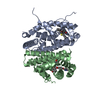
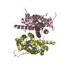
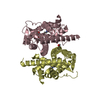
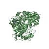
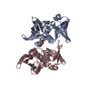
- PDB-5ex0: Crystal structure of human SMYD3 in complex with a MAP3K2 peptide -
+ Open data

- Basic information
| Entry | Database: PDB / ID: 5ex0 | |||||||||||||||||||||
|---|---|---|---|---|---|---|---|---|---|---|---|---|---|---|---|---|---|---|---|---|---|---|
| Title | Crystal structure of human SMYD3 in complex with a MAP3K2 peptide | |||||||||||||||||||||
 Components Components |
| |||||||||||||||||||||
 Keywords Keywords | TRANSFERASE / SET domain / methylation / chromatin / cancer | |||||||||||||||||||||
| Function / homology |  Function and homology information Function and homology informationhistone H4 methyltransferase activity / [histone H3]-lysine4 N-trimethyltransferase / histone H3K36 dimethyltransferase activity / myotube cell development / mitogen-activated protein kinase kinase kinase / histone H3K4 trimethyltransferase activity / RNA polymerase II intronic transcription regulatory region sequence-specific DNA binding / RNA polymerase II complex binding / cellular response to dexamethasone stimulus / MAP kinase kinase kinase activity ...histone H4 methyltransferase activity / [histone H3]-lysine4 N-trimethyltransferase / histone H3K36 dimethyltransferase activity / myotube cell development / mitogen-activated protein kinase kinase kinase / histone H3K4 trimethyltransferase activity / RNA polymerase II intronic transcription regulatory region sequence-specific DNA binding / RNA polymerase II complex binding / cellular response to dexamethasone stimulus / MAP kinase kinase kinase activity / establishment of protein localization / cellular response to mechanical stimulus / PKMTs methylate histone lysines / nucleosome assembly / methylation / protein kinase activity / intracellular signal transduction / RNA polymerase II cis-regulatory region sequence-specific DNA binding / protein serine kinase activity / protein serine/threonine kinase activity / protein kinase binding / positive regulation of transcription by RNA polymerase II / zinc ion binding / nucleoplasm / ATP binding / metal ion binding / nucleus / cytosol / cytoplasm Similarity search - Function | |||||||||||||||||||||
| Biological species |  Homo sapiens (human) Homo sapiens (human) | |||||||||||||||||||||
| Method |  X-RAY DIFFRACTION / SYNCHROTRON / MOLECULAR REPLACEMENT / Resolution: 2.7 Å X-RAY DIFFRACTION / SYNCHROTRON / MOLECULAR REPLACEMENT / Resolution: 2.7 Å | |||||||||||||||||||||
 Authors Authors | Fu, W. / Liu, N. / Qiao, Q. / Wang, M. / Min, J. / Zhu, B. / Xu, R.M. / Yang, N. | |||||||||||||||||||||
| Funding support |  China, 6items China, 6items
| |||||||||||||||||||||
 Citation Citation | Journal: J.Biol.Chem. / Year: 2016 Title: Structural Basis for Substrate Preference of SMYD3, a SET Domain-containing Protein Lysine Methyltransferase Authors: Fu, W. / Liu, N. / Qiao, Q. / Wang, M. / Min, J. / Zhu, B. / Xu, R.M. / Yang, N. | |||||||||||||||||||||
| History |
|
- Structure visualization
| Structure viewer | Molecule: MolmilJmol/JSmol |
|---|
- Downloads & links
-Download
| PDBx/mmCIF format | 5ex0.cif.gz | 108 KB | Display | PDBx/mmCIF format |
|---|---|---|---|---|
| PDB format | pdb5ex0.ent.gz | 78.5 KB | Display | PDB format |
| PDBx/mmJSON format | 5ex0.json.gz | Tree view | PDBx/mmJSON format | |
| Others |  Other downloads Other downloads |
-Validation report
| Arichive directory | https://data.pdbj.org/pub/pdb/validation_reports/ex/5ex0ftp://data.pdbj.org/pub/pdb/validation_reports/ex/5ex0 | HTTPS FTP |
|---|
-Related structure data
| Related structure data |  5ex3C 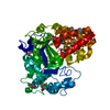 3mekS C: citing same article ( S: Starting model for refinement |
|---|---|
| Similar structure data |


-Links
 PDBj
PDBj


- Assembly
| Deposited unit | 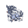
| ||||||||
|---|---|---|---|---|---|---|---|---|---|
| 1 |
| ||||||||
| Unit cell |
|
-Components
-Protein / Protein/peptide , 2 types, 2 molecules AD
| #1: Protein | Mass: 49541.492 Da / Num. of mol.: 1 / Mutation: K13N, K140R Source method: isolated from a genetically manipulated source Source: (gene. exp.) Homo sapiens (human) / Gene: SMYD3, ZMYND1, ZNFN3A1 / Plasmid: pET28a-smt / Production host:  References: UniProt: Q9H7B4, histone-lysine N-methyltransferase |
|---|---|
| #2: Protein/peptide | Mass: 1085.210 Da / Num. of mol.: 1 / Source method: obtained synthetically / Source: (synth.) Homo sapiens (human) / References: UniProt: Q9Y2U5*PLUS |
-Non-polymers , 4 types, 123 molecules 


| #3: Chemical |  Mass: 65.409 Da / Num. of mol.: 3 / Source method: obtained synthetically / Formula: Zn Mass: 65.409 Da / Num. of mol.: 3 / Source method: obtained synthetically / Formula: Zn#4: Chemical | ChemComp-SAH / |  Type: L-peptide linking / Mass: 384.411 Da / Num. of mol.: 1 / Source method: obtained synthetically / Formula: C14H20N6O5S Type: L-peptide linking / Mass: 384.411 Da / Num. of mol.: 1 / Source method: obtained synthetically / Formula: C14H20N6O5S#5: Chemical |  Mass: 60.052 Da / Num. of mol.: 2 / Source method: obtained synthetically / Formula: C2H4O2 Mass: 60.052 Da / Num. of mol.: 2 / Source method: obtained synthetically / Formula: C2H4O2#6: Water | ChemComp-HOH / | Mass: 18.015 Da / Num. of mol.: 117 / Source method: isolated from a natural source / Formula: H2O |
|---|
-Experimental details
-Experiment
| Experiment | Method: X-RAY DIFFRACTION / Number of used crystals: 1 |
|---|
- Sample preparation
| Crystal | Density Matthews: 3.25 Å3/Da / Density % sol: 62.2 % Description: THE ENTRY CONTAINS FRIEDEL PAIRS IN F_PLUS/MINUS COLUMNS. |
|---|---|
| Crystal grow | Temperature: 289 K / Method: vapor diffusion, hanging drop / pH: 7.5 / Details: 3.4M NaAC |
-Data collection
| Diffraction | Mean temperature: 100 K | |||||||||||||||||||||||||||||||||||||||||||||||||||||||||||||||||||||||||||||||||||||||||||||||||||
|---|---|---|---|---|---|---|---|---|---|---|---|---|---|---|---|---|---|---|---|---|---|---|---|---|---|---|---|---|---|---|---|---|---|---|---|---|---|---|---|---|---|---|---|---|---|---|---|---|---|---|---|---|---|---|---|---|---|---|---|---|---|---|---|---|---|---|---|---|---|---|---|---|---|---|---|---|---|---|---|---|---|---|---|---|---|---|---|---|---|---|---|---|---|---|---|---|---|---|---|---|
| Diffraction source | Source: SYNCHROTRON / Site: SSRF / Beamline: BL17U / Wavelength: 0.9791 Å | |||||||||||||||||||||||||||||||||||||||||||||||||||||||||||||||||||||||||||||||||||||||||||||||||||
| Detector | Type: ADSC QUANTUM 315r / Detector: CCD / Date: Dec 12, 2014 | |||||||||||||||||||||||||||||||||||||||||||||||||||||||||||||||||||||||||||||||||||||||||||||||||||
| Radiation | Monochromator: double crystal / Protocol: SINGLE WAVELENGTH / Monochromatic (M) / Laue (L): M / Scattering type: x-ray | |||||||||||||||||||||||||||||||||||||||||||||||||||||||||||||||||||||||||||||||||||||||||||||||||||
| Radiation wavelength | Wavelength: 0.9791 Å / Relative weight: 1 | |||||||||||||||||||||||||||||||||||||||||||||||||||||||||||||||||||||||||||||||||||||||||||||||||||
| Reflection | Resolution: 2.7→50 Å / Num. obs: 18908 / % possible obs: 99.9 % / Redundancy: 6 % / Biso Wilson estimate: 40.89 Å2 / Rmerge(I) obs: 0.111 / Rpim(I) all: 0.049 / Rrim(I) all: 0.121 / Χ2: 0.928 / Net I/av σ(I): 15.755 / Net I/σ(I): 7.4 / Num. measured all: 113209 | |||||||||||||||||||||||||||||||||||||||||||||||||||||||||||||||||||||||||||||||||||||||||||||||||||
| Reflection shell | Diffraction-ID: 1 / Rejects: _
|
- Processing
| Software |
| ||||||||||||||||||||||||||||||||||||||||||||||||||||||||
|---|---|---|---|---|---|---|---|---|---|---|---|---|---|---|---|---|---|---|---|---|---|---|---|---|---|---|---|---|---|---|---|---|---|---|---|---|---|---|---|---|---|---|---|---|---|---|---|---|---|---|---|---|---|---|---|---|---|
| Refinement | Method to determine structure: MOLECULAR REPLACEMENT Starting model: 3MEK Resolution: 2.7→47.66 Å / SU ML: 0.35 / Cross valid method: FREE R-VALUE / σ(F): 1.36 / Phase error: 22.55 / Stereochemistry target values: ML Details: SF FILE CONTAINS FRIEDEL PAIRS UNDER I/F_MINUS AND I/F_PLUS COLUMNS.
| ||||||||||||||||||||||||||||||||||||||||||||||||||||||||
| Solvent computation | Shrinkage radii: 0.9 Å / VDW probe radii: 1.11 Å / Solvent model: FLAT BULK SOLVENT MODEL | ||||||||||||||||||||||||||||||||||||||||||||||||||||||||
| Displacement parameters | Biso max: 85.53 Å2 / Biso mean: 36.4011 Å2 / Biso min: 17.9 Å2 | ||||||||||||||||||||||||||||||||||||||||||||||||||||||||
| Refinement step | Cycle: final / Resolution: 2.7→47.66 Å
| ||||||||||||||||||||||||||||||||||||||||||||||||||||||||
| Refine LS restraints |
| ||||||||||||||||||||||||||||||||||||||||||||||||||||||||
| LS refinement shell | Refine-ID: X-RAY DIFFRACTION / Total num. of bins used: 7
|
