 Movie
Movie Controller


+ Open data

- Basic information
| Entry | Database: PDB / ID: 5eur | |||||||||
|---|---|---|---|---|---|---|---|---|---|---|
| Title | Hypothetical protein SF216 from shigella flexneri 5a M90T | |||||||||
 Components Components | Uncharacterized protein | |||||||||
 Keywords Keywords | UNKNOWN FUNCTION / Shigella flexneri / Structural genomics / Glutathione S-transferase / Putative hot spot | |||||||||
| Function / homology | :  Function and homology information Function and homology information | |||||||||
| Biological species |  Shigella flexneri 5a str. M90T (bacteria) Shigella flexneri 5a str. M90T (bacteria) | |||||||||
| Method |  X-RAY DIFFRACTION / SYNCHROTRON / SAD / Resolution: 1.698 Å X-RAY DIFFRACTION / SYNCHROTRON / SAD / Resolution: 1.698 Å | |||||||||
 Authors Authors | Lee, Y.-S. / Seok, S.-H. / Kim, H.-N. / An, J.-G. / Chung, K.Y. / Won, H.-S. / Seo, M.-D. | |||||||||
| Funding support |  Korea, Republic Of, 2items Korea, Republic Of, 2items
| |||||||||
 Citation Citation | Journal: To Be Published Title: Crystal structure of SF216 from Shigella flexneri strain 5a Authors: Lee, Y.-S. / Seok, S.-H. / Kim, H.-N. / An, J.-G. / Chung, K.Y. / Won, H.-S. / Seo, M.-D. | |||||||||
| History |
|
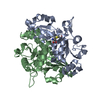
- Structure visualization
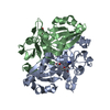
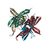
| Structure viewer | Molecule: MolmilJmol/JSmol |
|---|
- Downloads & links
-Download
| PDBx/mmCIF format | 5eur.cif.gz | 84.3 KB | Display | PDBx/mmCIF format |
|---|---|---|---|---|
| PDB format | pdb5eur.ent.gz | 63.7 KB | Display | PDB format |
| PDBx/mmJSON format | 5eur.json.gz | Tree view | PDBx/mmJSON format | |
| Others |  Other downloads Other downloads |
-Validation report
| Summary document | 5eur_validation.pdf.gz | 452.6 KB | Display | wwPDB validaton report |
|---|---|---|---|---|
| Full document | 5eur_full_validation.pdf.gz | 456 KB | Display | |
| Data in XML | 5eur_validation.xml.gz | 17.1 KB | Display | |
| Data in CIF | 5eur_validation.cif.gz | 24.4 KB | Display | |
| Arichive directory | https://data.pdbj.org/pub/pdb/validation_reports/eu/5eurftp://data.pdbj.org/pub/pdb/validation_reports/eu/5eur | HTTPS FTP |
-Related structure data
| Similar structure data |
|---|


-Links
 PDBj
PDBj- Assembly
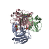
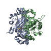
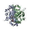
| Deposited unit | 
| ||||||||
|---|---|---|---|---|---|---|---|---|---|
| 1 |
| ||||||||
| Unit cell |
| ||||||||
| Components on special symmetry positions |
|
-Components
| #1: Protein | Mass: 15243.169 Da / Num. of mol.: 3 Source method: isolated from a genetically manipulated source Source: (gene. exp.) Shigella flexneri 5a str. M90T (bacteria)Gene: SF5M90T_216 / Plasmid: pET21a (+) / Production host: #2: Chemical | ChemComp-MG / |   Mass: 24.305 Da / Num. of mol.: 1 / Source method: obtained synthetically / Formula: Mg Mass: 24.305 Da / Num. of mol.: 1 / Source method: obtained synthetically / Formula: Mg#3: Chemical | ChemComp-SO4 / |   Mass: 96.063 Da / Num. of mol.: 1 / Source method: obtained synthetically / Formula: SO4 Mass: 96.063 Da / Num. of mol.: 1 / Source method: obtained synthetically / Formula: SO4#4: Water | ChemComp-HOH / |  Mass: 18.015 Da / Num. of mol.: 279 / Source method: isolated from a natural source / Formula: H2O Mass: 18.015 Da / Num. of mol.: 279 / Source method: isolated from a natural source / Formula: H2O |
|---|
-Experimental details
-Experiment
| Experiment | Method: X-RAY DIFFRACTION / Number of used crystals: 1 |
|---|
- Sample preparation
| Crystal | Density Matthews: 2.23 Å3/Da / Density % sol: 47.31 % Description: hexahedron in shape, with two trapezoidal faces |
|---|---|
| Crystal grow | Temperature: 293 K / Method: vapor diffusion, sitting drop Details: 0.1 M sodium HEPES, 2 M ammonium sulfate, 2% v/v PEG 400 |
-Data collection
| Diffraction | Mean temperature: 100 K |
|---|---|
| Diffraction source | Source: SYNCHROTRON / Site: PAL/PLS / Beamline: 7A (6B, 6C1) / Wavelength: 0.9797 Å |
| Detector | Type: ADSC QUANTUM 270 / Detector: CCD / Date: Sep 30, 2014 |
| Radiation | Protocol: SINGLE WAVELENGTH / Monochromatic (M) / Laue (L): M / Scattering type: x-ray |
| Radiation wavelength | Wavelength: 0.9797 Å / Relative weight: 1 |
| Reflection | Resolution: 1.698→50 Å / Num. obs: 44562 / % possible obs: 95.1 % / Redundancy: 4.8 % / Biso Wilson estimate: 19.13 Å2 / Rmerge(I) obs: 0.013 / Net I/σ(I): 36.9 |
| Reflection shell | Resolution: 1.7→1.76 Å / Redundancy: 3.9 % / Rmerge(I) obs: 0.322 / Mean I/σ(I) obs: 6.2 / % possible all: 88.4 |
- Processing
| Software |
| |||||||||||||||||||||||||||||||||||||||||||||||||||||||||||||||||||||||||||||||||||||||||||||||||||||||||
|---|---|---|---|---|---|---|---|---|---|---|---|---|---|---|---|---|---|---|---|---|---|---|---|---|---|---|---|---|---|---|---|---|---|---|---|---|---|---|---|---|---|---|---|---|---|---|---|---|---|---|---|---|---|---|---|---|---|---|---|---|---|---|---|---|---|---|---|---|---|---|---|---|---|---|---|---|---|---|---|---|---|---|---|---|---|---|---|---|---|---|---|---|---|---|---|---|---|---|---|---|---|---|---|---|---|---|
| Refinement | Method to determine structure: SAD / Resolution: 1.698→24.478 Å / SU ML: 0.18 / Cross valid method: FREE R-VALUE / σ(F): 1.34 / Phase error: 22.92 / Stereochemistry target values: ML
| |||||||||||||||||||||||||||||||||||||||||||||||||||||||||||||||||||||||||||||||||||||||||||||||||||||||||
| Solvent computation | Shrinkage radii: 0.9 Å / VDW probe radii: 1.11 Å / Solvent model: FLAT BULK SOLVENT MODEL | |||||||||||||||||||||||||||||||||||||||||||||||||||||||||||||||||||||||||||||||||||||||||||||||||||||||||
| Refinement step | Cycle: LAST / Resolution: 1.698→24.478 Å
| |||||||||||||||||||||||||||||||||||||||||||||||||||||||||||||||||||||||||||||||||||||||||||||||||||||||||
| Refine LS restraints |
| |||||||||||||||||||||||||||||||||||||||||||||||||||||||||||||||||||||||||||||||||||||||||||||||||||||||||
| LS refinement shell |
|
