 Movie
Movie Controller

[English] 日本語
 Yorodumi
- PDB-5esp: Crystal Structure of LAGLIDADG Meganuclease I-PanMI with coordina... -
+ Open data

- Basic information
| Entry | Database: PDB / ID: 5esp | ||||||
|---|---|---|---|---|---|---|---|
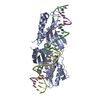
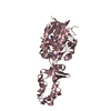
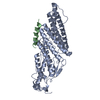
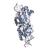
| Title | Crystal Structure of LAGLIDADG Meganuclease I-PanMI with coordinated Calcium ions | ||||||
 Components Components |
| ||||||
 Keywords Keywords | HYDROLASE/DNA / Hydrolase-DNA complex / LAGLIDADG / homing endonuclease / meganuclease | ||||||
| Function / homology |  Function and homology information Function and homology information | ||||||
| Biological species |  Podospora anserina (fungus) Podospora anserina (fungus)synthetic construct (others) | ||||||
| Method |  X-RAY DIFFRACTION / MOLECULAR REPLACEMENT / Resolution: 2.995 Å X-RAY DIFFRACTION / MOLECULAR REPLACEMENT / Resolution: 2.995 Å | ||||||
 Authors Authors | Hallinan, J.P. / Stoddard, B.L. | ||||||
| Funding support |  United States, 1items United States, 1items
| ||||||
 Citation Citation | Journal: Structure / Year: 2016 Title: Indirect DNA Sequence Recognition and Its Impact on Nuclease Cleavage Activity. Authors: Lambert, A.R. / Hallinan, J.P. / Shen, B.W. / Chik, J.K. / Bolduc, J.M. / Kulshina, N. / Robins, L.I. / Kaiser, B.K. / Jarjour, J. / Havens, K. / Scharenberg, A.M. / Stoddard, B.L. | ||||||
| History |
|
- Structure visualization
| Structure viewer | Molecule: MolmilJmol/JSmol |
|---|
- Downloads & links
-Download
| PDBx/mmCIF format | 5esp.cif.gz | 184 KB | Display | PDBx/mmCIF format |
|---|---|---|---|---|
| PDB format | pdb5esp.ent.gz | 138.3 KB | Display | PDB format |
| PDBx/mmJSON format | 5esp.json.gz | Tree view | PDBx/mmJSON format | |
| Others |  Other downloads Other downloads |
-Validation report
| Arichive directory | https://data.pdbj.org/pub/pdb/validation_reports/es/5espftp://data.pdbj.org/pub/pdb/validation_reports/es/5esp | HTTPS FTP |
|---|
-Related structure data
| Related structure data |  4yhxC  4yisC  4yitC  4z1zC  4z20C  4jee S: Starting model for refinement C: citing same article ( |
|---|---|
| Similar structure data |


-Links
 PDBj
PDBj


- Assembly
| Deposited unit | 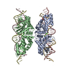
| ||||||||
|---|---|---|---|---|---|---|---|---|---|
| 1 | 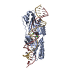
| ||||||||
| 2 | 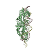
| ||||||||
| Unit cell |
|
-Components
-Protein , 1 types, 2 molecules AB
| #1: Protein | Mass: 33995.645 Da / Num. of mol.: 2 / Fragment: UNP residues 136-433 Source method: isolated from a genetically manipulated source Source: (gene. exp.) Podospora anserina (fungus) / Strain: S / ATCC MYA-4624 / DSM 980 / FGSC 10383 / Production host:  |
|---|
-DNA chain , 2 types, 4 molecules GCHD
| #2: DNA chain | Mass: 8092.227 Da / Num. of mol.: 2 / Source method: obtained synthetically / Source: (synth.) synthetic construct (others) #3: DNA chain | Mass: 8501.480 Da / Num. of mol.: 2 / Source method: obtained synthetically / Source: (synth.) synthetic construct (others) |
|---|
-Non-polymers , 3 types, 8 molecules 


| #4: Chemical | ChemComp-CA /  Mass: 40.078 Da / Num. of mol.: 4 / Source method: obtained synthetically / Formula: Ca Mass: 40.078 Da / Num. of mol.: 4 / Source method: obtained synthetically / Formula: Ca#5: Chemical |  Mass: 92.094 Da / Num. of mol.: 2 / Source method: obtained synthetically / Formula: C3H8O3 Mass: 92.094 Da / Num. of mol.: 2 / Source method: obtained synthetically / Formula: C3H8O3#6: Water | ChemComp-HOH / | Mass: 18.015 Da / Num. of mol.: 2 / Source method: isolated from a natural source / Formula: H2O |
|---|
-Experimental details
-Experiment
| Experiment | Method: X-RAY DIFFRACTION / Number of used crystals: 1 |
|---|
- Sample preparation
| Crystal | Density Matthews: 2.76 Å3/Da / Density % sol: 55.51 % |
|---|---|
| Crystal grow | Temperature: 298 K / Method: vapor diffusion, hanging drop / pH: 7 / Details: 28% PEG 3350, 5mM Calcium Chloride |
-Data collection
| Diffraction | Mean temperature: 100 K | |||||||||||||||||||||||||||||||||||||||||||||||||||||||||||||||||||||||||||||||||||||||||||||||||||
|---|---|---|---|---|---|---|---|---|---|---|---|---|---|---|---|---|---|---|---|---|---|---|---|---|---|---|---|---|---|---|---|---|---|---|---|---|---|---|---|---|---|---|---|---|---|---|---|---|---|---|---|---|---|---|---|---|---|---|---|---|---|---|---|---|---|---|---|---|---|---|---|---|---|---|---|---|---|---|---|---|---|---|---|---|---|---|---|---|---|---|---|---|---|---|---|---|---|---|---|---|
| Diffraction source | Source: ROTATING ANODE / Type: RIGAKU MICROMAX-007 HF / Wavelength: 1.54 Å | |||||||||||||||||||||||||||||||||||||||||||||||||||||||||||||||||||||||||||||||||||||||||||||||||||
| Detector | Type: RIGAKU SATURN 944+ / Detector: CCD / Date: Sep 10, 2015 | |||||||||||||||||||||||||||||||||||||||||||||||||||||||||||||||||||||||||||||||||||||||||||||||||||
| Radiation | Monochromator: VARIMAX HF OPTICS / Protocol: SINGLE WAVELENGTH / Monochromatic (M) / Laue (L): M / Scattering type: x-ray | |||||||||||||||||||||||||||||||||||||||||||||||||||||||||||||||||||||||||||||||||||||||||||||||||||
| Radiation wavelength | Wavelength: 1.54 Å / Relative weight: 1 | |||||||||||||||||||||||||||||||||||||||||||||||||||||||||||||||||||||||||||||||||||||||||||||||||||
| Reflection | Resolution: 2.995→30.969 Å / Num. obs: 21733 / % possible obs: 98.6 % / Redundancy: 5.8 % / Rmerge(I) obs: 0.157 / Rpim(I) all: 0.07 / Rrim(I) all: 0.173 / Χ2: 1.518 / Net I/av σ(I): 13.226 / Net I/σ(I): 7.7 / Num. measured all: 127010 | |||||||||||||||||||||||||||||||||||||||||||||||||||||||||||||||||||||||||||||||||||||||||||||||||||
| Reflection shell | Diffraction-ID: 1 / Rejects: _
|
- Processing
| Software |
| ||||||||||||||||||||||||
|---|---|---|---|---|---|---|---|---|---|---|---|---|---|---|---|---|---|---|---|---|---|---|---|---|---|
| Refinement | Method to determine structure: MOLECULAR REPLACEMENT Starting model: 4JEE 4jee Resolution: 2.995→30.969 Å / SU ML: 0.46 / Cross valid method: FREE R-VALUE / σ(F): 1.34 / Phase error: 28.53 / Stereochemistry target values: ML
| ||||||||||||||||||||||||
| Solvent computation | Shrinkage radii: 0.9 Å / VDW probe radii: 1.11 Å / Solvent model: FLAT BULK SOLVENT MODEL | ||||||||||||||||||||||||
| Displacement parameters | Biso max: 227.68 Å2 / Biso mean: 56.0929 Å2 / Biso min: 10.41 Å2 | ||||||||||||||||||||||||
| Refinement step | Cycle: final / Resolution: 2.995→30.969 Å
|
