| 登録情報 | データベース: PDB / ID: 5er5
|
|---|
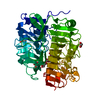
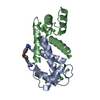
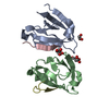
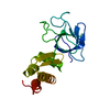
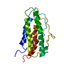
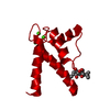
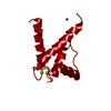
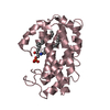
| タイトル | Crystal Structure of Calcium-loaded S100B bound to SC1990 |
|---|
 要素 要素 | Protein S100-B |
|---|
 キーワード キーワード | METAL BINDING PROTEIN/INHIBITOR / malignant melanoma / calcium binding / complex / covalent inhibitor / METAL BINDING PROTEIN-INHIBITOR complex |
|---|
| 機能・相同性 |  機能・相同性情報 機能・相同性情報
Advanced glycosylation endproduct receptor signaling / TRAF6 mediated NF-kB activation / TAK1-dependent IKK and NF-kappa-B activation / adaptive thermogenesis / kinase inhibitor activity / sympathetic neuron projection extension / positive regulation of complement activation / RAGE receptor binding / negative regulation of monocyte chemotactic protein-1 production / S100 protein binding ...Advanced glycosylation endproduct receptor signaling / TRAF6 mediated NF-kB activation / TAK1-dependent IKK and NF-kappa-B activation / adaptive thermogenesis / kinase inhibitor activity / sympathetic neuron projection extension / positive regulation of complement activation / RAGE receptor binding / negative regulation of monocyte chemotactic protein-1 production / S100 protein binding / phosphorylation / regulation of neuronal synaptic plasticity / ruffle / positive regulation of neuron differentiation / astrocyte activation / axonogenesis / tau protein binding / memory / calcium-dependent protein binding / regulation of translation / learning or memory / positive regulation of canonical NF-kappaB signal transduction / cell adhesion / ciliary basal body / neuronal cell body / positive regulation of cell population proliferation / calcium ion binding / perinuclear region of cytoplasm / protein homodimerization activity / extracellular space / extracellular region / zinc ion binding / nucleoplasm / nucleus / cytosol / cytoplasm類似検索 - 分子機能 Protein S100-B / S-100/ICaBP type calcium binding protein signature. / S100/Calcium binding protein 7/8-like, conserved site / S100/CaBP-9k-type, calcium binding, subdomain / S-100/ICaBP type calcium binding domain / S-100/ICaBP type calcium binding domain / EF hand domain / EF-hand / Recoverin; domain 1 / EF-hand, calcium binding motif ...Protein S100-B / S-100/ICaBP type calcium binding protein signature. / S100/Calcium binding protein 7/8-like, conserved site / S100/CaBP-9k-type, calcium binding, subdomain / S-100/ICaBP type calcium binding domain / S-100/ICaBP type calcium binding domain / EF hand domain / EF-hand / Recoverin; domain 1 / EF-hand, calcium binding motif / EF-Hand 1, calcium-binding site / EF-hand calcium-binding domain. / EF-hand calcium-binding domain profile. / EF-hand domain / EF-hand domain pair / Orthogonal Bundle / Mainly Alpha類似検索 - ドメイン・相同性 |
|---|
| 生物種 |   Bos taurus (ウシ) Bos taurus (ウシ) |
|---|
| 手法 |  X線回折 / シンクロトロン / 分子置換 / 解像度: 1.26 Å X線回折 / シンクロトロン / 分子置換 / 解像度: 1.26 Å |
|---|
 データ登録者 データ登録者 | Cavalier, M.C. / Melville, Z.E. / Aligholizadeh, E. / Fang, L. / Alasady, M.J. / Pierce, A.D. / Wilder, P.T. / MacKerell Jr., A.D. / Weber, D.J. |
|---|
| 資金援助 |  米国, 5件 米国, 5件 | 組織 | 認可番号 | 国 |
|---|
| National Institutes of Health/National Cancer Institute (NIH/NCI) | CA107331 | 米国 | | National Institutes of Health/National Institute of General Medical Sciences (NIH/NIGMS) | GM58888 | 米国 | | National Institutes of Health/National Cancer Institute (NIH/NCI) | CA154274 | 米国 | | National Institutes of Health/National Institute of Arthritis and Musculoskeletal and Skin Diseases (NIH/NIAMS) | AR007592 | 米国 | | National Institutes of Health/National Institute of General Medical Sciences (NIH/NIGMS) | P41GM103393 | 米国 |
|
|---|
 引用 引用 | ジャーナル: Acta Crystallogr D Struct Biol / 年: 2016
タイトル: Novel protein-inhibitor interactions in site 3 of Ca(2+)-bound S100B as discovered by X-ray crystallography.
著者: Cavalier, M.C. / Melville, Z. / Aligholizadeh, E. / Raman, E.P. / Yu, W. / Fang, L. / Alasady, M. / Pierce, A.D. / Wilder, P.T. / MacKerell, A.D. / Weber, D.J. |
|---|
| 履歴 | | 登録 | 2015年11月13日 | 登録サイト: RCSB / 処理サイト: RCSB |
|---|
| 改定 1.0 | 2016年6月8日 | Provider: repository / タイプ: Initial release |
|---|
| 改定 1.1 | 2017年9月27日 | Group: Author supporting evidence / Derived calculations / カテゴリ: pdbx_audit_support / pdbx_struct_oper_list
Item: _pdbx_audit_support.funding_organization / _pdbx_struct_oper_list.symmetry_operation |
|---|
| 改定 1.2 | 2018年4月18日 | Group: Data collection / Database references / カテゴリ: citation / citation_author
Item: _citation.country / _citation.journal_abbrev ..._citation.country / _citation.journal_abbrev / _citation.journal_id_ASTM / _citation.journal_id_ISSN / _citation.journal_volume / _citation.pdbx_database_id_PubMed / _citation.title |
|---|
| 改定 1.3 | 2019年12月4日 | Group: Author supporting evidence / カテゴリ: pdbx_audit_support / Item: _pdbx_audit_support.funding_organization |
|---|
| 改定 1.4 | 2023年9月27日 | Group: Data collection / Database references / Refinement description
カテゴリ: chem_comp_atom / chem_comp_bond ...chem_comp_atom / chem_comp_bond / database_2 / pdbx_initial_refinement_model
Item: _database_2.pdbx_DOI / _database_2.pdbx_database_accession |
|---|
|
|---|
 ムービー
ムービー コントローラー


 データを開く
 基本情報
基本情報 構造の表示
構造の表示 ダウンロードとリンク
 その他のダウンロード
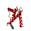


 PDBj
PDBj 集合体
集合体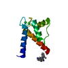


 分子量: 40.078 Da / 分子数: 2 / 由来タイプ: 合成 / 式: Ca
 分子量: 314.404 Da / 分子数: 1 / 由来タイプ: 合成 / 式: C21H20N3
 分子量: 18.015 Da / 分子数: 158 / 由来タイプ: 天然 / 式: H2O
 試料調製
試料調製 解析
解析