[English] 日本語
Yorodumi
- PDB-5eq9: Crystal structure of Medicago truncatula Histidinol-Phosphate Pho... -

+
Open data


ID or keywords:

Loading...

-
Basic information

Entry
Database: PDB / ID: 5eq9
TitleCrystal structure of Medicago truncatula Histidinol-Phosphate Phosphatase (MtHPP) in complex with L-histidinol phosphate and Mg2+
ComponentsInositol monophosphatase
KeywordsBIOSYNTHETIC PROTEIN / histidine biosynthesis / metabolic pathways / dimer / plant
Function / homology
Function and homology information


histidinol-phosphatase / histidinol-phosphatase activity / L-histidine biosynthetic process / metal ion binding
Similarity search - Function
Histidinol-phosphate phosphatase, putative, inositol monophosphatase / Inositol monophosphatase, metal-binding site / Inositol monophosphatase family signature 1. / Inositol monophosphatase-like / Inositol monophosphatase family / D-Maltodextrin-Binding Protein; domain 2 - #80 / Fructose-1,6-Bisphosphatase, subunit A, domain 1 / Fructose-1,6-Bisphosphatase; Chain A, domain 1 / D-Maltodextrin-Binding Protein; domain 2 / 2-Layer Sandwich ...Histidinol-phosphate phosphatase, putative, inositol monophosphatase / Inositol monophosphatase, metal-binding site / Inositol monophosphatase family signature 1. / Inositol monophosphatase-like / Inositol monophosphatase family / D-Maltodextrin-Binding Protein; domain 2 - #80 / Fructose-1,6-Bisphosphatase, subunit A, domain 1 / Fructose-1,6-Bisphosphatase; Chain A, domain 1 / D-Maltodextrin-Binding Protein; domain 2 / 2-Layer Sandwich / 3-Layer(aba) Sandwich / Alpha Beta
Similarity search - Domain/homology
Chem-HSA / histidinol-phosphatase
Similarity search - Component
Biological speciesMedicago truncatula (barrel medic)
MethodX-RAY DIFFRACTION / SYNCHROTRON / MOLECULAR REPLACEMENT / Resolution: 1.36 Å
AuthorsRuszkowski, M. / Dauter, Z.
Citation
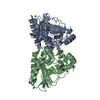
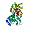
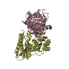
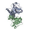
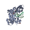
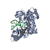
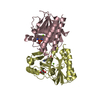
Journal: J.Biol.Chem. / Year: 2016
Title: Structural Studies of Medicago truncatula Histidinol Phosphate Phosphatase from Inositol Monophosphatase Superfamily Reveal Details of Penultimate Step of Histidine Biosynthesis in Plants.
Authors: Ruszkowski, M. / Dauter, Z.
History
DepositionNov 12, 2015Deposition site: RCSB / Processing site: RCSB
Revision 1.0Mar 30, 2016Provider: repository / Type: Initial release
Revision 1.1Jun 1, 2016Group: Database references
Revision 1.2Mar 6, 2024Group: Data collection / Database references / Derived calculations
Category: chem_comp_atom / chem_comp_bond ...chem_comp_atom / chem_comp_bond / citation / database_2 / pdbx_struct_oper_list
Item: _citation.journal_id_CSD / _database_2.pdbx_DOI ..._citation.journal_id_CSD / _database_2.pdbx_DOI / _database_2.pdbx_database_accession / _pdbx_struct_oper_list.symmetry_operation

-
Structure visualization

Structure viewerMolecule:
MolmilJmol/JSmol

Downloads & links

-
Assembly

Deposited unit
A: Inositol monophosphatase
B: Inositol monophosphatase
C: Inositol monophosphatase
D: Inositol monophosphatase
hetero molecules


Theoretical massNumber of molelcules
Total (without water)122,71113
Polymers121,6374
Non-polymers1,0749
Water23,6901315
1
A: Inositol monophosphatase
B: Inositol monophosphatase
hetero molecules


Theoretical massNumber of molelcules
Total (without water)61,3096
Polymers60,8192
Non-polymers4914
Water362
TypeNameSymmetry operationNumber
identity operation1_555x,y,z1
Buried area3610 Å2
ΔGint-29 kcal/mol
Surface area20300 Å2
MethodPISA
2
C: Inositol monophosphatase
D: Inositol monophosphatase
hetero molecules


Theoretical massNumber of molelcules
Total (without water)61,4027
Polymers60,8192
Non-polymers5835
Water362
TypeNameSymmetry operationNumber
identity operation1_555x,y,z1
Buried area3520 Å2
ΔGint-29 kcal/mol
Surface area20400 Å2
MethodPISA
Unit cell
Length a, b, c (Å)61.885, 89.802, 92.593
Angle α, β, γ (deg.)90.00, 97.07, 90.00
Int Tables number4
Space group name H-MP1211

-
Components

#1: Protein
Inositol monophosphatase


Mass: 30409.285 Da / Num. of mol.: 4
Source method: isolated from a genetically manipulated source
Source: (gene. exp.) Medicago truncatula (barrel medic) / Gene: MTR_3g117220 / Plasmid: pMCSG68 / Production host: Escherichia coli (E. coli) / Strain (production host): BL21 Gold / References: UniProt: G7J7Q5
#2: Chemical
ChemComp-HSA / PHOSPHORIC ACID MONO-[2-AMINO-3-(3H-IMIDAZOL-4-YL)-PROPYL]ESTER


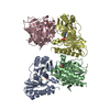
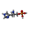
Mass: 221.151 Da / Num. of mol.: 4 / Source method: obtained synthetically / Formula: C6H12N3O4P
#3: Chemical
ChemComp-MG / MAGNESIUM ION


Mass: 24.305 Da / Num. of mol.: 4 / Source method: obtained synthetically / Formula: Mg
#4: Chemical ChemComp-GOL / GLYCEROL / GLYCERIN / PROPANE-1,2,3-TRIOL


Mass: 92.094 Da / Num. of mol.: 1 / Source method: obtained synthetically / Formula: C3H8O3
#5: Water ChemComp-HOH / water


Mass: 18.015 Da / Num. of mol.: 1315 / Source method: isolated from a natural source / Formula: H2O

-
Experimental details

-
Experiment

ExperimentMethod: X-RAY DIFFRACTION

-
Sample preparation

CrystalDensity Matthews: 2.1 Å3/Da / Density % sol: 41.4 %
Crystal growTemperature: 292 K / Method: vapor diffusion, hanging drop / pH: 8
Details: Protein at 19 mg/ml concentration. 15% PEG 3350, 0.2 M diammonium hydrogen phosphate, pH 8.0. Crystal washed in a crystallization solution supplemented with 2 mM MgCl2, 5 mM HOLP and 20% ...Details: Protein at 19 mg/ml concentration. 15% PEG 3350, 0.2 M diammonium hydrogen phosphate, pH 8.0. Crystal washed in a crystallization solution supplemented with 2 mM MgCl2, 5 mM HOLP and 20% glycerol and immediately vitrified in liquid nitrogen

-
Data collection

DiffractionMean temperature: 100 K
Diffraction sourceSource: SYNCHROTRON / Site: APS / Beamline: 19-ID / Wavelength: 0.9793 Å
DetectorType: ADSC QUANTUM 315r / Detector: CCD / Date: Jul 30, 2015
RadiationProtocol: SINGLE WAVELENGTH / Monochromatic (M) / Laue (L): M / Scattering type: x-ray
Radiation wavelengthWavelength: 0.9793 Å / Relative weight: 1
ReflectionResolution: 1.36→40 Å / Num. obs: 214644 / % possible obs: 99.8 % / Redundancy: 5.1 % / Rmerge(I) obs: 0.072 / Net I/σ(I): 15
Reflection shellResolution: 1.36→1.44 Å / Redundancy: 5 % / Rmerge(I) obs: 0.82 / Mean I/σ(I) obs: 1.9 / % possible all: 98.8

-
Processing

Software
NameVersionClassification
REFMAC5.8.0131refinement
XDSdata reduction
XDSdata scaling
PHASERphasing
RefinementMethod to determine structure: MOLECULAR REPLACEMENT / Resolution: 1.36→36.25 Å / Cor.coef. Fo:Fc: 0.983 / Cor.coef. Fo:Fc free: 0.975 / SU B: 1.922 / SU ML: 0.033 / Cross valid method: FREE R-VALUE / ESU R: 0.043 / ESU R Free: 0.045 / Stereochemistry target values: MAXIMUM LIKELIHOOD / Details: HYDROGENS HAVE BEEN ADDED IN THE RIDING POSITIONS
RfactorNum. reflection% reflectionSelection details
Rfree0.156 1074 0.5 %RANDOM
Rwork0.116 ---
obs0.116 213570 99.8 %-
Solvent computationIon probe radii: 0.8 Å / Shrinkage radii: 0.8 Å / VDW probe radii: 1.2 Å / Solvent model: MASK
Displacement parametersBiso mean: 17.576 Å2
Baniso -1Baniso -2Baniso -3
1--0.18 Å20 Å20.81 Å2
2--0.04 Å20 Å2
3----0.06 Å2
Refinement stepCycle: 1 / Resolution: 1.36→36.25 Å
ProteinNucleic acidLigandSolventTotal
Num. atoms7967 0 66 1315 9348
Refine LS restraints
Refine-IDTypeDev idealDev ideal targetNumber
X-RAY DIFFRACTIONr_bond_refined_d0.020.0198262
X-RAY DIFFRACTIONr_bond_other_d0.0020.027817
X-RAY DIFFRACTIONr_angle_refined_deg1.91.95811259
X-RAY DIFFRACTIONr_angle_other_deg1.112317996
X-RAY DIFFRACTIONr_dihedral_angle_1_deg6.29351040
X-RAY DIFFRACTIONr_dihedral_angle_2_deg36.72924.507355
X-RAY DIFFRACTIONr_dihedral_angle_3_deg13.056151331
X-RAY DIFFRACTIONr_dihedral_angle_4_deg14.1611536
X-RAY DIFFRACTIONr_chiral_restr0.1370.21272
X-RAY DIFFRACTIONr_gen_planes_refined0.0090.0219365
X-RAY DIFFRACTIONr_gen_planes_other0.0020.021889
X-RAY DIFFRACTIONr_nbd_refined
X-RAY DIFFRACTIONr_nbd_other
X-RAY DIFFRACTIONr_nbtor_refined
X-RAY DIFFRACTIONr_nbtor_other
X-RAY DIFFRACTIONr_xyhbond_nbd_refined
X-RAY DIFFRACTIONr_xyhbond_nbd_other
X-RAY DIFFRACTIONr_metal_ion_refined
X-RAY DIFFRACTIONr_metal_ion_other
X-RAY DIFFRACTIONr_symmetry_vdw_refined
X-RAY DIFFRACTIONr_symmetry_vdw_other
X-RAY DIFFRACTIONr_symmetry_hbond_refined
X-RAY DIFFRACTIONr_symmetry_hbond_other
X-RAY DIFFRACTIONr_symmetry_metal_ion_refined
X-RAY DIFFRACTIONr_symmetry_metal_ion_other
X-RAY DIFFRACTIONr_mcbond_it2.2961.3264126
X-RAY DIFFRACTIONr_mcbond_other2.2961.3264126
X-RAY DIFFRACTIONr_mcangle_it2.9091.9935143
X-RAY DIFFRACTIONr_mcangle_other2.9091.9935144
X-RAY DIFFRACTIONr_scbond_it3.9491.7694136
X-RAY DIFFRACTIONr_scbond_other3.9481.7694136
X-RAY DIFFRACTIONr_scangle_it
X-RAY DIFFRACTIONr_scangle_other4.7692.4846106
X-RAY DIFFRACTIONr_long_range_B_refined5.78413.78210537
X-RAY DIFFRACTIONr_long_range_B_other5.78413.78310538
X-RAY DIFFRACTIONr_rigid_bond_restr3.702316079
X-RAY DIFFRACTIONr_sphericity_free39.4995303
X-RAY DIFFRACTIONr_sphericity_bonded14.752516921
LS refinement shellResolution: 1.36→1.4 Å
RfactorNum. reflection% reflection
Rfree0.346 78 -
Rwork0.265 15365 -
obs--97.6 %

+
About Yorodumi

-
News

-
Feb 9, 2022. New format data for meta-information of EMDB entries

New format data for meta-information of EMDB entries

  • Version 3 of the EMDB header file is now the official format.
  • The previous official version 1.9 will be removed from the archive.

Related info.:EMDB header

External links:wwPDB to switch to version 3 of the EMDB data model

-
Aug 12, 2020. Covid-19 info

Covid-19 info

URL: https://pdbjlvh1.pdbj.org/emnavi/covid19.php

New page: Covid-19 featured information page in EM Navigator.

Related info.:Covid-19 info / Mar 5, 2020. Novel coronavirus structure data

+
Mar 5, 2020. Novel coronavirus structure data

Novel coronavirus structure data

Related info.:Yorodumi Speices / Aug 12, 2020. Covid-19 info

External links:COVID-19 featured content - PDBj / Molecule of the Month (242):Coronavirus Proteases

+
Jan 31, 2019. EMDB accession codes are about to change! (news from PDBe EMDB page)

EMDB accession codes are about to change! (news from PDBe EMDB page)

  • The allocation of 4 digits for EMDB accession codes will soon come to an end. Whilst these codes will remain in use, new EMDB accession codes will include an additional digit and will expand incrementally as the available range of codes is exhausted. The current 4-digit format prefixed with “EMD-” (i.e. EMD-XXXX) will advance to a 5-digit format (i.e. EMD-XXXXX), and so on. It is currently estimated that the 4-digit codes will be depleted around Spring 2019, at which point the 5-digit format will come into force.
  • The EM Navigator/Yorodumi systems omit the EMD- prefix.

Related info.:Q: What is EMD? / ID/Accession-code notation in Yorodumi/EM Navigator

External links:EMDB Accession Codes are Changing Soon! / Contact to PDBj

+
Jul 12, 2017. Major update of PDB

Major update of PDB

  • wwPDB released updated PDB data conforming to the new PDBx/mmCIF dictionary.
  • This is a major update changing the version number from 4 to 5, and with Remediation, in which all the entries are updated.
  • In this update, many items about electron microscopy experimental information are reorganized (e.g. em_software).
  • Now, EM Navigator and Yorodumi are based on the updated data.

External links:wwPDB Remediation / Enriched Model Files Conforming to OneDep Data Standards Now Available in the PDB FTP Archive

-
Yorodumi

Thousand views of thousand structures

  • Yorodumi is a browser for structure data from EMDB, PDB, SASBDB, etc.
  • This page is also the successor to EM Navigator detail page, and also detail information page/front-end page for Omokage search.
  • The word "yorodu" (or yorozu) is an old Japanese word meaning "ten thousand". "mi" (miru) is to see.

Related info.:EMDB / PDB / SASBDB / Comparison of 3 databanks / Yorodumi Search / Aug 31, 2016. New EM Navigator & Yorodumi / Yorodumi Papers / Jmol/JSmol / Function and homology information / Changes in new EM Navigator and Yorodumi

Read more