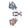
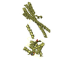
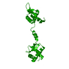
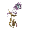
[English] 日本語
Yorodumi
- PDB-5en6: Crystal structure of the Smu1-RED complex (SeMet) of Caenorhabdit... -

+
Open data


ID or keywords:

Loading...

-
Basic information

Entry
Database: PDB / ID: 5en6
TitleCrystal structure of the Smu1-RED complex (SeMet) of Caenorhabditis elegans
Components
  • SMU-1
  • Suppressor of Mec and Unc defects
KeywordsSPLICING / LisH motif / CTLH / dimer / heterotetramer / splicing factor
Function / homology
Function and homology information


nematode larval development / muscle organ morphogenesis / mechanosensory behavior / locomotion / U2-type precatalytic spliceosome / embryo development ending in birth or egg hatching / precatalytic spliceosome / regulation of alternative mRNA splicing, via spliceosome / neuron development / RNA splicing ...nematode larval development / muscle organ morphogenesis / mechanosensory behavior / locomotion / U2-type precatalytic spliceosome / embryo development ending in birth or egg hatching / precatalytic spliceosome / regulation of alternative mRNA splicing, via spliceosome / neuron development / RNA splicing / spliceosomal complex / mRNA splicing, via spliceosome / nucleolus / protein homodimerization activity / identical protein binding / nucleus
Similarity search - Function
Protein RED, C-terminal / RED-like, N-terminal / Protein Red-like / RED-like protein C-terminal region / RED-like protein N-terminal region / WD40 repeat-containing protein SMU1 / LisH-like dimerisation domain / C-terminal to LisH motif. / CTLH, C-terminal LisH motif / C-terminal to LisH (CTLH) motif profile. ...Protein RED, C-terminal / RED-like, N-terminal / Protein Red-like / RED-like protein C-terminal region / RED-like protein N-terminal region / WD40 repeat-containing protein SMU1 / LisH-like dimerisation domain / C-terminal to LisH motif. / CTLH, C-terminal LisH motif / C-terminal to LisH (CTLH) motif profile. / Lissencephaly type-1-like homology motif / LIS1 homology (LisH) motif profile. / LIS1 homology motif / G-protein beta WD-40 repeat / WD domain, G-beta repeat / WD40 repeats / WD40 repeat / Trp-Asp (WD) repeats profile. / Trp-Asp (WD) repeats circular profile. / WD40-repeat-containing domain superfamily / WD40/YVTN repeat-like-containing domain superfamily
Similarity search - Domain/homology
Smu-1 suppressor of mec-8 and unc-52 protein / Smu-2 suppressor of mec-8 and unc-52 protein
Similarity search - Component
Biological speciesCaenorhabditis elegans (invertebrata)
MethodX-RAY DIFFRACTION / SYNCHROTRON / MAD / molecular replacement / Resolution: 3.103 Å
AuthorsUlrich, A.K.C. / Wahl, M.C.
Funding support Germany, 1items
OrganizationGrant numberCountry
DFGWA1126/7-1 Germany
CitationJournal: to be published
Title: Crystal structure of the Smu1-RED complex (SeMet) of Caenorhabditis elegans
Authors: Ulrich, A.K.C. / Schulz, J.F. / Kamprad, A. / Schuetze, T. / Wahl, M.C.
History
DepositionNov 9, 2015Deposition site: RCSB / Processing site: PDBE
Revision 1.0Apr 20, 2016Provider: repository / Type: Initial release

-
Structure visualization

Structure viewerMolecule:
MolmilJmol/JSmol

Downloads & links

-
Assembly

Deposited unit
A: SMU-1
B: SMU-1
C: Suppressor of Mec and Unc defects
D: Suppressor of Mec and Unc defects


Theoretical massNumber of molelcules
Total (without water)55,6284
Polymers55,6284
Non-polymers00
Water1,00956
1
A: SMU-1
D: Suppressor of Mec and Unc defects

A: SMU-1
D: Suppressor of Mec and Unc defects


Theoretical massNumber of molelcules
Total (without water)55,6284
Polymers55,6284
Non-polymers00
Water362
TypeNameSymmetry operationNumber
identity operation1_555x,y,z1
crystal symmetry operation3_455-x-1,y,-z+1/21
Buried area4930 Å2
ΔGint-47 kcal/mol
Surface area22580 Å2
MethodPISA
2
B: SMU-1
C: Suppressor of Mec and Unc defects

B: SMU-1
C: Suppressor of Mec and Unc defects


Theoretical massNumber of molelcules
Total (without water)55,6284
Polymers55,6284
Non-polymers00
Water724
TypeNameSymmetry operationNumber
identity operation1_555x,y,z1
crystal symmetry operation3_353-x-2,y,-z-3/21
Buried area5020 Å2
ΔGint-48 kcal/mol
Surface area22070 Å2
MethodPISA
Unit cell
Length a, b, c (Å)166.669, 183.443, 41.078
Angle α, β, γ (deg.)90.000, 90.000, 90.000
Int Tables number20
Space group name H-MC2221
Noncrystallographic symmetry (NCS)NCS domain:
IDEns-IDDetails
11chain A
21chain B

NCS domain segments:
Dom-IDComponent-IDEns-IDSelection detailsAuth asym-IDAuth seq-ID
111chain AA5 - 180
211chain BB7 - 175

-
Components

#1: Protein SMU-1 / Suppressor of Mec and Unc defects


Mass: 20802.508 Da / Num. of mol.: 2 / Fragment: NTR, UNP residues 2-181
Source method: isolated from a genetically manipulated source
Source: (gene. exp.) Caenorhabditis elegans (invertebrata) / Gene: smu-1, CC4.3, CELE_CC4.3 / Plasmid: pETM11 / Production host: Escherichia coli BL21(DE3) (bacteria) / Variant (production host): RIL / References: UniProt: G5EEG7
#2: Protein Suppressor of Mec and Unc defects


Mass: 7011.264 Da / Num. of mol.: 2 / Fragment: UNP residues 163-223
Source method: isolated from a genetically manipulated source
Source: (gene. exp.) Caenorhabditis elegans (invertebrata) / Gene: smu-2, CELE_Y49F6B.4, Y49F6B.4 / Plasmid: pETM11 / Production host: Escherichia coli BL21(DE3) (bacteria) / Variant (production host): RIL / References: UniProt: Q9N4U5
#3: Water ChemComp-HOH / water / Water


Mass: 18.015 Da / Num. of mol.: 56 / Source method: isolated from a natural source / Formula: H2O

-
Experimental details

-
Experiment

ExperimentMethod: X-RAY DIFFRACTION / Number of used crystals: 1

-
Sample preparation

CrystalDensity Matthews: 2.82 Å3/Da / Density % sol: 56.36 %
Crystal growTemperature: 291.15 K / Method: vapor diffusion
Details: 0.1 M HEPES, pH 7.0, 0.02 M MgCl2, 19 % (w/v) polyacrilic acid 5,100
PH range: 7

-
Data collection

DiffractionMean temperature: 100 K
Diffraction sourceSource: SYNCHROTRON / Site: BESSY / Beamline: 14.1 / Wavelength: 0.979531, 0.979771, 0.977427
DetectorType: DECTRIS PILATUS 6M / Detector: PIXEL / Date: Jul 14, 2015
RadiationProtocol: MAD / Monochromatic (M) / Laue (L): M / Scattering type: x-ray
Radiation wavelength
IDWavelength (Å)Relative weight
10.9771
20.981
30.9795311
40.9797711
50.9774271
ReflectionResolution: 3.092→45.86 Å / Num. obs: 11889 / % possible obs: 99.7 % / Redundancy: 12.8 % / Biso Wilson estimate: 102.6 Å2 / Rsym value: 0.103 / Net I/σ(I): 19.83
Reflection shellResolution: 3.1→3.29 Å / Redundancy: 12.2 % / Rmerge(I) obs: 1.283 / Mean I/σ(I) obs: 2.04 / % possible all: 99.2

-
Phasing

Phasing
Method
MAD
molecular replacement

-
Processing

Software
NameVersionClassification
PHENIXrefinement
XSCALEdata scaling
SHARPphasing
PDB_EXTRACT3.15data extraction
XDSdata reduction
RefinementMethod to determine structure: MAD / Resolution: 3.103→45.861 Å / SU ML: 0.46 / Cross valid method: FREE R-VALUE / σ(F): 1.36 / Phase error: 37.32 / Stereochemistry target values: MLHL
RfactorNum. reflection% reflectionSelection details
Rfree0.3206 561 4.72 %Random selection
Rwork0.2831 11320 --
obs0.2849 11881 99.66 %-
Solvent computationShrinkage radii: 0.9 Å / VDW probe radii: 1.11 Å / Solvent model: FLAT BULK SOLVENT MODEL
Displacement parametersBiso max: 197.03 Å2 / Biso mean: 111.2601 Å2 / Biso min: 41.99 Å2
Refinement stepCycle: final / Resolution: 3.103→45.861 Å
ProteinNucleic acidLigandSolventTotal
Num. atoms3130 0 0 56 3186
Biso mean---91.93 -
Num. residues----393
Refine LS restraints
Refine-IDTypeDev idealNumber
X-RAY DIFFRACTIONf_bond_d0.0033174
X-RAY DIFFRACTIONf_angle_d0.7224293
X-RAY DIFFRACTIONf_chiral_restr0.028513
X-RAY DIFFRACTIONf_plane_restr0.002548
X-RAY DIFFRACTIONf_dihedral_angle_d14.0471203
Refine LS restraints NCS
Ens-IDDom-IDAuth asym-IDNumberRefine-IDRmsType
11A1407X-RAY DIFFRACTION6.296TORSIONAL
12B1407X-RAY DIFFRACTION6.296TORSIONAL
LS refinement shell

Refine-ID: X-RAY DIFFRACTION / Total num. of bins used: 4

Resolution (Å)Rfactor RfreeNum. reflection RfreeRfactor RworkNum. reflection RworkNum. reflection all% reflection obs (%)
3.103-3.41520.39021400.36642753289399
3.4152-3.90910.38061420.331527702912100
3.9091-4.92420.33881270.278628292956100
4.9242-45.86570.28061520.255829683120100

+
About Yorodumi

-
News

-
Feb 9, 2022. New format data for meta-information of EMDB entries

New format data for meta-information of EMDB entries

  • Version 3 of the EMDB header file is now the official format.
  • The previous official version 1.9 will be removed from the archive.

Related info.:EMDB header

External links:wwPDB to switch to version 3 of the EMDB data model

-
Aug 12, 2020. Covid-19 info

Covid-19 info

URL: https://pdbj.org/emnavi/covid19.php

New page: Covid-19 featured information page in EM Navigator.

Related info.:Covid-19 info / Mar 5, 2020. Novel coronavirus structure data

+
Mar 5, 2020. Novel coronavirus structure data

Novel coronavirus structure data

Related info.:Yorodumi Speices / Aug 12, 2020. Covid-19 info

External links:COVID-19 featured content - PDBj / Molecule of the Month (242):Coronavirus Proteases

+
Jan 31, 2019. EMDB accession codes are about to change! (news from PDBe EMDB page)

EMDB accession codes are about to change! (news from PDBe EMDB page)

  • The allocation of 4 digits for EMDB accession codes will soon come to an end. Whilst these codes will remain in use, new EMDB accession codes will include an additional digit and will expand incrementally as the available range of codes is exhausted. The current 4-digit format prefixed with “EMD-” (i.e. EMD-XXXX) will advance to a 5-digit format (i.e. EMD-XXXXX), and so on. It is currently estimated that the 4-digit codes will be depleted around Spring 2019, at which point the 5-digit format will come into force.
  • The EM Navigator/Yorodumi systems omit the EMD- prefix.

Related info.:Q: What is EMD? / ID/Accession-code notation in Yorodumi/EM Navigator

External links:EMDB Accession Codes are Changing Soon! / Contact to PDBj

+
Jul 12, 2017. Major update of PDB

Major update of PDB

  • wwPDB released updated PDB data conforming to the new PDBx/mmCIF dictionary.
  • This is a major update changing the version number from 4 to 5, and with Remediation, in which all the entries are updated.
  • In this update, many items about electron microscopy experimental information are reorganized (e.g. em_software).
  • Now, EM Navigator and Yorodumi are based on the updated data.

External links:wwPDB Remediation / Enriched Model Files Conforming to OneDep Data Standards Now Available in the PDB FTP Archive

-
Yorodumi

Thousand views of thousand structures

  • Yorodumi is a browser for structure data from EMDB, PDB, SASBDB, etc.
  • This page is also the successor to EM Navigator detail page, and also detail information page/front-end page for Omokage search.
  • The word "yorodu" (or yorozu) is an old Japanese word meaning "ten thousand". "mi" (miru) is to see.

Related info.:EMDB / PDB / SASBDB / Comparison of 3 databanks / Yorodumi Search / Aug 31, 2016. New EM Navigator & Yorodumi / Yorodumi Papers / Jmol/JSmol / Function and homology information / Changes in new EM Navigator and Yorodumi

Read more