 Movie
Movie Controller

[English] 日本語
 Yorodumi
- PDB-5em3: Crystal structure of the human BRPF1 bromodomain in complex with SEED9 -
+ Open data

- Basic information
| Entry | Database: PDB / ID: 5em3 | |||||||||
|---|---|---|---|---|---|---|---|---|---|---|
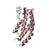
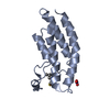
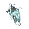
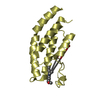
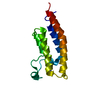
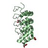
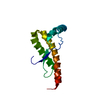
| Title | Crystal structure of the human BRPF1 bromodomain in complex with SEED9 | |||||||||
 Components Components | Peregrin | |||||||||
 Keywords Keywords | DNA BINDING PROTEIN / Bromodomain and PHD finger-containing protein 1(BRPF1) / monocytic leukemia zinc-finger (MOZ) / Inhibitor / transcription | |||||||||
| Function / homology |  Function and homology information Function and homology informationacetyltransferase activator activity / MOZ/MORF histone acetyltransferase complex / regulation of developmental process / regulation of hemopoiesis / histone acetyltransferase complex / Regulation of TP53 Activity through Acetylation / HATs acetylate histones / chromatin remodeling / regulation of transcription by RNA polymerase II / regulation of DNA-templated transcription ...acetyltransferase activator activity / MOZ/MORF histone acetyltransferase complex / regulation of developmental process / regulation of hemopoiesis / histone acetyltransferase complex / Regulation of TP53 Activity through Acetylation / HATs acetylate histones / chromatin remodeling / regulation of transcription by RNA polymerase II / regulation of DNA-templated transcription / positive regulation of DNA-templated transcription / DNA binding / zinc ion binding / nucleoplasm / nucleus / plasma membrane / cytoplasm / cytosol Similarity search - Function | |||||||||
| Biological species |  Homo sapiens (human) Homo sapiens (human) | |||||||||
| Method |  X-RAY DIFFRACTION / SYNCHROTRON / MOLECULAR REPLACEMENT / Resolution: 1.4 Å X-RAY DIFFRACTION / SYNCHROTRON / MOLECULAR REPLACEMENT / Resolution: 1.4 Å | |||||||||
 Authors Authors | Zhu, J. / Caflisch, A. | |||||||||
| Funding support |  Switzerland, 1items Switzerland, 1items
| |||||||||
 Citation Citation | Journal: J.Med.Chem. / Year: 2016 Title: Twenty Crystal Structures of Bromodomain and PHD Finger Containing Protein 1 (BRPF1)/Ligand Complexes Reveal Conserved Binding Motifs and Rare Interactions. Authors: Zhu, J. / Caflisch, A. | |||||||||
| History |
|
- Structure visualization
| Structure viewer | Molecule: MolmilJmol/JSmol |
|---|
- Downloads & links
-Download
| PDBx/mmCIF format | 5em3.cif.gz | 71.4 KB | Display | PDBx/mmCIF format |
|---|---|---|---|---|
| PDB format | pdb5em3.ent.gz | 52.9 KB | Display | PDB format |
| PDBx/mmJSON format | 5em3.json.gz | Tree view | PDBx/mmJSON format | |
| Others |  Other downloads Other downloads |
-Validation report
| Arichive directory | https://data.pdbj.org/pub/pdb/validation_reports/em/5em3ftp://data.pdbj.org/pub/pdb/validation_reports/em/5em3 | HTTPS FTP |
|---|
-Related structure data
| Related structure data |  5c7nC 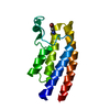 5c85C 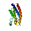 5c87C  5dy7C 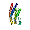 5dyaC 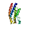 5dycC 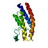 5e3dC 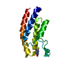 5e3gC 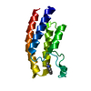 5eprC  5epsC 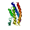 5eq1C 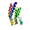 5etbC 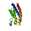 5etdC 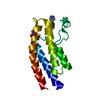 5ev9C 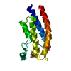 5evaC 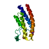 5ewcC 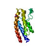 5ewdC 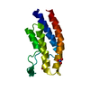 5ewhC 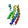 4lc2S C: citing same article ( S: Starting model for refinement |
|---|---|
| Similar structure data |


-Links
 PDBj
PDBj


- Assembly
| Deposited unit | 
| |||||||||
|---|---|---|---|---|---|---|---|---|---|---|
| 1 |
| |||||||||
| Unit cell |
| |||||||||
| Components on special symmetry positions |
|
-Components
| #1: Protein | Mass: 13703.698 Da / Num. of mol.: 1 / Fragment: UNP residues 626-740 Source method: isolated from a genetically manipulated source Source: (gene. exp.) Homo sapiens (human) / Gene: BRPF1, BR140 / Production host:  | ||||
|---|---|---|---|---|---|
| #2: Chemical |   Mass: 145.158 Da / Num. of mol.: 2 / Source method: obtained synthetically / Formula: C9H7NO Mass: 145.158 Da / Num. of mol.: 2 / Source method: obtained synthetically / Formula: C9H7NO#3: Chemical |   Mass: 62.005 Da / Num. of mol.: 2 / Source method: obtained synthetically / Formula: NO3 Mass: 62.005 Da / Num. of mol.: 2 / Source method: obtained synthetically / Formula: NO3#4: Water | ChemComp-HOH / |  Mass: 18.015 Da / Num. of mol.: 170 / Source method: isolated from a natural source / Formula: H2O Mass: 18.015 Da / Num. of mol.: 170 / Source method: isolated from a natural source / Formula: H2O |
-Experimental details
-Experiment
| Experiment | Method: X-RAY DIFFRACTION / Number of used crystals: 1 |
|---|
- Sample preparation
| Crystal | Density Matthews: 2.41 Å3/Da / Density % sol: 48.93 % |
|---|---|
| Crystal grow | Temperature: 277 K / Method: vapor diffusion, hanging drop Details: 0.1 M Sodium-Acetate, pH5.5, 0.15 M Sodium Nitrate, 20% PEG3350 |
-Data collection
| Diffraction | Mean temperature: 100 K |
|---|---|
| Diffraction source | Source: SYNCHROTRON / Site: SLS / Beamline: X06SA / Wavelength: 0.99987 Å |
| Detector | Type: DECTRIS PILATUS 6M / Detector: PIXEL / Date: Jul 5, 2015 |
| Radiation | Protocol: SINGLE WAVELENGTH / Monochromatic (M) / Laue (L): M / Scattering type: x-ray |
| Radiation wavelength | Wavelength: 0.99987 Å / Relative weight: 1 |
| Reflection | Resolution: 1.4→40.2 Å / Num. obs: 26409 / % possible obs: 99.7 % / Redundancy: 9.2 % / Rmerge(I) obs: 0.049 / Net I/σ(I): 25.4 |
| Reflection shell | Resolution: 1.4→1.48 Å / Redundancy: 8.8 % / Rmerge(I) obs: 0.236 / Mean I/σ(I) obs: 8.4 |
- Processing
| Software |
| ||||||||||||||||||||||||||||||||||||||||||||||||||||||||||||||||||||||
|---|---|---|---|---|---|---|---|---|---|---|---|---|---|---|---|---|---|---|---|---|---|---|---|---|---|---|---|---|---|---|---|---|---|---|---|---|---|---|---|---|---|---|---|---|---|---|---|---|---|---|---|---|---|---|---|---|---|---|---|---|---|---|---|---|---|---|---|---|---|---|---|
| Refinement | Method to determine structure: MOLECULAR REPLACEMENT Starting model: 4LC2 Resolution: 1.4→40.198 Å / SU ML: 0.13 / Cross valid method: THROUGHOUT / σ(F): 1.37 / Phase error: 18.4 / Stereochemistry target values: ML
| ||||||||||||||||||||||||||||||||||||||||||||||||||||||||||||||||||||||
| Solvent computation | Shrinkage radii: 0.9 Å / VDW probe radii: 1.11 Å / Solvent model: FLAT BULK SOLVENT MODEL | ||||||||||||||||||||||||||||||||||||||||||||||||||||||||||||||||||||||
| Refinement step | Cycle: LAST / Resolution: 1.4→40.198 Å
| ||||||||||||||||||||||||||||||||||||||||||||||||||||||||||||||||||||||
| Refine LS restraints |
| ||||||||||||||||||||||||||||||||||||||||||||||||||||||||||||||||||||||
| LS refinement shell |
| ||||||||||||||||||||||||||||||||||||||||||||||||||||||||||||||||||||||
| Refinement TLS params. | Method: refined / Origin x: 27.3639 Å / Origin y: 1.1575 Å / Origin z: -10.0924 Å
| ||||||||||||||||||||||||||||||||||||||||||||||||||||||||||||||||||||||
| Refinement TLS group | Selection details: all |
