 Movie
Movie Controller

[English] 日本語
 Yorodumi
- PDB-5ejy: Structure of Dictyostelium Discoideum Myosin VII MyTH4-FERM MF1 domain -
+ Open data

- Basic information
| Entry | Database: PDB / ID: 5ejy | ||||||
|---|---|---|---|---|---|---|---|
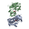
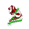
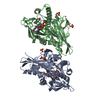
| Title | Structure of Dictyostelium Discoideum Myosin VII MyTH4-FERM MF1 domain | ||||||
 Components Components | Myosin-I heavy chain | ||||||
 Keywords Keywords | MOTOR PROTEIN / Molecular motor / myosin / myosin tail / MyTH4-FERM | ||||||
| Function / homology |  Function and homology information Function and homology informationspore germination / filopodium tip / actin filament-based movement / filopodium assembly / myosin complex / microfilament motor activity / cell-substrate adhesion / cell leading edge / phagocytosis / phagocytic cup ...spore germination / filopodium tip / actin filament-based movement / filopodium assembly / myosin complex / microfilament motor activity / cell-substrate adhesion / cell leading edge / phagocytosis / phagocytic cup / actin filament organization / filopodium / response to calcium ion / cell morphogenesis / endocytosis / actin filament binding / actin cytoskeleton / cell cortex / microtubule binding / ATP binding / membrane / cytoplasm / cytosol Similarity search - Function | ||||||
| Biological species |  | ||||||
| Method |  X-RAY DIFFRACTION / SYNCHROTRON / MOLECULAR REPLACEMENT / Resolution: 1.9 Å X-RAY DIFFRACTION / SYNCHROTRON / MOLECULAR REPLACEMENT / Resolution: 1.9 Å | ||||||
 Authors Authors | Sirigu, S. / Titus, M.A. / Houdusse, A. | ||||||
 Citation Citation | Journal: Proc.Natl.Acad.Sci.USA / Year: 2016 Title: Myosin MyTH4-FERM structures highlight important principles of convergent evolution. Authors: Planelles-Herrero, V.J. / Blanc, F. / Sirigu, S. / Sirkia, H. / Clause, J. / Sourigues, Y. / Johnsrud, D.O. / Amigues, B. / Cecchini, M. / Gilbert, S.P. / Houdusse, A. / Titus, M.A. | ||||||
| History |
|
- Structure visualization
| Structure viewer | Molecule: MolmilJmol/JSmol |
|---|
- Downloads & links
-Download
| PDBx/mmCIF format | 5ejy.cif.gz | 131.2 KB | Display | PDBx/mmCIF format |
|---|---|---|---|---|
| PDB format | pdb5ejy.ent.gz | 95 KB | Display | PDB format |
| PDBx/mmJSON format | 5ejy.json.gz | Tree view | PDBx/mmJSON format | |
| Others |  Other downloads Other downloads |
-Validation report
| Arichive directory | https://data.pdbj.org/pub/pdb/validation_reports/ej/5ejyftp://data.pdbj.org/pub/pdb/validation_reports/ej/5ejy | HTTPS FTP |
|---|
-Related structure data
| Related structure data |  5ejqC  5ejrC  5ejsC 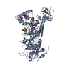 3pvlS S: Starting model for refinement C: citing same article ( |
|---|---|
| Similar structure data |


-Links
 PDBj
PDBj


- Assembly
| Deposited unit | 
| ||||||||
|---|---|---|---|---|---|---|---|---|---|
| 1 |
| ||||||||
| Unit cell |
|
-Components
| #1: Protein | Mass: 57437.789 Da / Num. of mol.: 1 Source method: isolated from a genetically manipulated source Source: (gene. exp.)  | ||||
|---|---|---|---|---|---|
| #2: Chemical |   Mass: 35.453 Da / Num. of mol.: 2 / Source method: obtained synthetically / Formula: Cl Mass: 35.453 Da / Num. of mol.: 2 / Source method: obtained synthetically / Formula: Cl#3: Chemical |   Mass: 1529.829 Da / Num. of mol.: 2 / Source method: obtained synthetically / Formula: C69H140O35 / Comment: precipitant*YM Mass: 1529.829 Da / Num. of mol.: 2 / Source method: obtained synthetically / Formula: C69H140O35 / Comment: precipitant*YM#4: Water | ChemComp-HOH / |  Mass: 18.015 Da / Num. of mol.: 503 / Source method: isolated from a natural source / Formula: H2O Mass: 18.015 Da / Num. of mol.: 503 / Source method: isolated from a natural source / Formula: H2O |
-Experimental details
-Experiment
| Experiment | Method: X-RAY DIFFRACTION / Number of used crystals: 1 |
|---|
- Sample preparation
| Crystal | Density Matthews: 3.53 Å3/Da / Density % sol: 65.14 % |
|---|---|
| Crystal grow | Temperature: 290 K / Method: vapor diffusion, hanging drop / pH: 6 / Details: 18% PEG 8000, 100 mM MES pH 6.0 |
-Data collection
| Diffraction | Mean temperature: 100 K |
|---|---|
| Diffraction source | Source: SYNCHROTRON / Site: SOLEIL  / Beamline: PROXIMA 2 / Wavelength: 0.97242 Å / Beamline: PROXIMA 2 / Wavelength: 0.97242 Å |
| Detector | Type: ADSC QUANTUM 315r / Detector: CCD / Date: Mar 16, 2014 |
| Radiation | Protocol: SINGLE WAVELENGTH / Monochromatic (M) / Laue (L): M / Scattering type: x-ray |
| Radiation wavelength | Wavelength: 0.97242 Å / Relative weight: 1 |
| Reflection | Resolution: 1.9→50 Å / Num. obs: 69695 / % possible obs: 99.3 % / Redundancy: 5.9 % / Rsym value: 0.057 / Net I/σ(I): 18.36 |
| Reflection shell | Resolution: 1.9→1.96 Å / Redundancy: 5.14 % / Mean I/σ(I) obs: 2.27 / % possible all: 97.1 |
- Processing
| Software |
| |||||||||||||||||||||||||||||||||||||||||||||||||||||||||||||||||||||||||||||
|---|---|---|---|---|---|---|---|---|---|---|---|---|---|---|---|---|---|---|---|---|---|---|---|---|---|---|---|---|---|---|---|---|---|---|---|---|---|---|---|---|---|---|---|---|---|---|---|---|---|---|---|---|---|---|---|---|---|---|---|---|---|---|---|---|---|---|---|---|---|---|---|---|---|---|---|---|---|---|
| Refinement | Method to determine structure: MOLECULAR REPLACEMENT Starting model: 3pvl Resolution: 1.9→43.308 Å / FOM work R set: 0.8307 / SU ML: 0.23 / Cross valid method: FREE R-VALUE / σ(F): 2 / Phase error: 23.67 / Stereochemistry target values: ML
| |||||||||||||||||||||||||||||||||||||||||||||||||||||||||||||||||||||||||||||
| Solvent computation | Shrinkage radii: 0.83 Å / VDW probe radii: 1.1 Å / Solvent model: FLAT BULK SOLVENT MODEL / Bsol: 62.354 Å2 / ksol: 0.357 e/Å3 | |||||||||||||||||||||||||||||||||||||||||||||||||||||||||||||||||||||||||||||
| Displacement parameters | Biso max: 81.32 Å2 / Biso mean: 36.31 Å2 / Biso min: 16.22 Å2
| |||||||||||||||||||||||||||||||||||||||||||||||||||||||||||||||||||||||||||||
| Refinement step | Cycle: final / Resolution: 1.9→43.308 Å
| |||||||||||||||||||||||||||||||||||||||||||||||||||||||||||||||||||||||||||||
| Refine LS restraints |
| |||||||||||||||||||||||||||||||||||||||||||||||||||||||||||||||||||||||||||||
| LS refinement shell | Refine-ID: X-RAY DIFFRACTION / Total num. of bins used: 10
|
