 Movie
Movie Controller

[English] 日本語
 Yorodumi
- PDB-5eec: Crystal structure of KPC-2 beta-lactamase in complex with the S02... -
+ Open data

- Basic information
| Entry | Database: PDB / ID: 5eec | ||||||
|---|---|---|---|---|---|---|---|
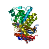
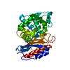
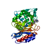
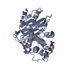
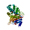
| Title | Crystal structure of KPC-2 beta-lactamase in complex with the S02030 boronic acid inhibitor | ||||||
 Components Components | Carbapenem-hydrolyzing beta-lactamase KPC | ||||||
 Keywords Keywords | HYDROLASE/HYDROLASE INHIBITOR / transition state inhibitor / HYDROLASE-HYDROLASE INHIBITOR complex | ||||||
| Function / homology |  Function and homology information Function and homology informationAntimicrobial resistance / beta-lactam antibiotic catabolic process / beta-lactamase activity / beta-lactamase / response to antibiotic / cytosol Similarity search - Function | ||||||
| Biological species |  Klebsiella pneumoniae (bacteria) Klebsiella pneumoniae (bacteria) | ||||||
| Method |  X-RAY DIFFRACTION / Resolution: 1.87 Å X-RAY DIFFRACTION / Resolution: 1.87 Å | ||||||
 Authors Authors | Nguyen, N.Q. / van den Akker, F. | ||||||
 Citation Citation | Journal: Antimicrob.Agents Chemother. / Year: 2016 Title: Crystal Structures of KPC-2 and SHV-1 beta-Lactamases in Complex with the Boronic Acid Transition State Analog S02030. Authors: Nguyen, N.Q. / Krishnan, N.P. / Rojas, L.J. / Prati, F. / Caselli, E. / Romagnoli, C. / Bonomo, R.A. / van den Akker, F. | ||||||
| History |
|
- Structure visualization
| Structure viewer | Molecule: MolmilJmol/JSmol |
|---|
- Downloads & links
-Download
| PDBx/mmCIF format | 5eec.cif.gz | 124.2 KB | Display | PDBx/mmCIF format |
|---|---|---|---|---|
| PDB format | pdb5eec.ent.gz | 94.5 KB | Display | PDB format |
| PDBx/mmJSON format | 5eec.json.gz | Tree view | PDBx/mmJSON format | |
| Others |  Other downloads Other downloads |
-Validation report
| Arichive directory | https://data.pdbj.org/pub/pdb/validation_reports/ee/5eecftp://data.pdbj.org/pub/pdb/validation_reports/ee/5eec | HTTPS FTP |
|---|
-Related structure data


-Links
 PDBj
PDBj


- Assembly
| Deposited unit | 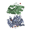
| ||||||||
|---|---|---|---|---|---|---|---|---|---|
| 1 | 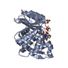
| ||||||||
| 2 | 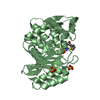
| ||||||||
| Unit cell |
|
-Components
| #1: Protein | Mass: 28504.074 Da / Num. of mol.: 2 Source method: isolated from a genetically manipulated source Source: (gene. exp.) Klebsiella pneumoniae (bacteria) / Gene: bla, kpc, kpc1 / Production host: #2: Chemical |   Mass: 324.121 Da / Num. of mol.: 2 / Source method: obtained synthetically / Formula: C11H13BN4O5S Mass: 324.121 Da / Num. of mol.: 2 / Source method: obtained synthetically / Formula: C11H13BN4O5S#3: Chemical | ChemComp-SO4 /   Mass: 96.063 Da / Num. of mol.: 6 / Source method: obtained synthetically / Formula: SO4 Mass: 96.063 Da / Num. of mol.: 6 / Source method: obtained synthetically / Formula: SO4#4: Water | ChemComp-HOH / |  Mass: 18.015 Da / Num. of mol.: 419 / Source method: isolated from a natural source / Formula: H2O Mass: 18.015 Da / Num. of mol.: 419 / Source method: isolated from a natural source / Formula: H2OHas protein modification | Y | |
|---|
-Experimental details
-Experiment
| Experiment | Method: X-RAY DIFFRACTION / Number of used crystals: 1 |
|---|
- Sample preparation
| Crystal | Density Matthews: 2.2 Å3/Da / Density % sol: 44.2 % |
|---|---|
| Crystal grow | Temperature: 293 K / Method: vapor diffusion, sitting drop / pH: 4.5 Details: 30% PEG 8000, 0.2 M lithium sulfate, and 0.1 M sodium acetate (pH 4.5) and 10:1 molar ratio with S02030 inhibitor |
-Data collection
| Diffraction | Mean temperature: 100 K | ||||||||||||||||||||||||||||||||||||||||||||||||||||||||||||||||||
|---|---|---|---|---|---|---|---|---|---|---|---|---|---|---|---|---|---|---|---|---|---|---|---|---|---|---|---|---|---|---|---|---|---|---|---|---|---|---|---|---|---|---|---|---|---|---|---|---|---|---|---|---|---|---|---|---|---|---|---|---|---|---|---|---|---|---|---|
| Diffraction source | Source: ROTATING ANODE / Type: RIGAKU MICROMAX-007 / Wavelength: 1.542 Å | ||||||||||||||||||||||||||||||||||||||||||||||||||||||||||||||||||
| Detector | Type: RIGAKU SATURN 944+ / Detector: CCD / Date: Oct 22, 2014 | ||||||||||||||||||||||||||||||||||||||||||||||||||||||||||||||||||
| Radiation | Monochromator: Cu filter / Protocol: SINGLE WAVELENGTH / Monochromatic (M) / Laue (L): M / Scattering type: x-ray | ||||||||||||||||||||||||||||||||||||||||||||||||||||||||||||||||||
| Radiation wavelength | Wavelength: 1.542 Å / Relative weight: 1 | ||||||||||||||||||||||||||||||||||||||||||||||||||||||||||||||||||
| Reflection | Resolution: 1.85→50 Å / Num. obs: 39177 / % possible obs: 96.7 % / Redundancy: 2.9 % / Rmerge(I) obs: 0.107 / Χ2: 1.179 / Net I/av σ(I): 9.005 / Net I/σ(I): 9.7 / Num. measured all: 113565 | ||||||||||||||||||||||||||||||||||||||||||||||||||||||||||||||||||
| Reflection shell | Diffraction-ID: 1 / Rejects: _
|
- Processing
| Software |
| ||||||||||||||||||||||||||||||||||||||||||||||||||||||||||||
|---|---|---|---|---|---|---|---|---|---|---|---|---|---|---|---|---|---|---|---|---|---|---|---|---|---|---|---|---|---|---|---|---|---|---|---|---|---|---|---|---|---|---|---|---|---|---|---|---|---|---|---|---|---|---|---|---|---|---|---|---|---|
| Refinement | Resolution: 1.87→32.88 Å / Cor.coef. Fo:Fc: 0.948 / Cor.coef. Fo:Fc free: 0.92 / SU B: 3.211 / SU ML: 0.094 / Cross valid method: THROUGHOUT / σ(F): 0 / ESU R: 0.155 / ESU R Free: 0.138 / Stereochemistry target values: MAXIMUM LIKELIHOOD Details: HYDROGENS HAVE BEEN ADDED IN THE RIDING POSITIONS U VALUES : REFINED INDIVIDUALLY
| ||||||||||||||||||||||||||||||||||||||||||||||||||||||||||||
| Solvent computation | Ion probe radii: 0.8 Å / Shrinkage radii: 0.8 Å / VDW probe radii: 1.2 Å / Solvent model: MASK | ||||||||||||||||||||||||||||||||||||||||||||||||||||||||||||
| Displacement parameters | Biso max: 500 Å2 / Biso mean: 13.044 Å2 / Biso min: 2.95 Å2
| ||||||||||||||||||||||||||||||||||||||||||||||||||||||||||||
| Refinement step | Cycle: final / Resolution: 1.87→32.88 Å
| ||||||||||||||||||||||||||||||||||||||||||||||||||||||||||||
| Refine LS restraints |
| ||||||||||||||||||||||||||||||||||||||||||||||||||||||||||||
| LS refinement shell | Resolution: 1.866→1.915 Å / Total num. of bins used: 20
|
