 Movie
Movie Controller

[English] 日本語
 Yorodumi
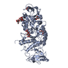
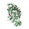
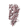
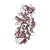
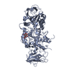
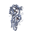
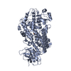
- PDB-5e6z: Crystal structure of Ecoli Branching Enzyme with beta cyclodextrin -
+ Open data

- Basic information
| Entry | Database: PDB / ID: 5e6z | |||||||||
|---|---|---|---|---|---|---|---|---|---|---|
| Title | Crystal structure of Ecoli Branching Enzyme with beta cyclodextrin | |||||||||
 Components Components | 1,4-alpha-glucan branching enzyme GlgB | |||||||||
 Keywords Keywords | TRANSFERASE / Branching Enzyme / Cyclodextrin / Glycogen / Starch / glucan | |||||||||
| Function / homology |  Function and homology information Function and homology informationcation binding / 1,4-alpha-glucan branching enzyme / 1,4-alpha-glucan branching enzyme activity / glycogen biosynthetic process / hydrolase activity, hydrolyzing O-glycosyl compounds / cytosol Similarity search - Function | |||||||||
| Biological species |  | |||||||||
| Method |  X-RAY DIFFRACTION / SYNCHROTRON / MOLECULAR REPLACEMENT / Resolution: 1.878 Å X-RAY DIFFRACTION / SYNCHROTRON / MOLECULAR REPLACEMENT / Resolution: 1.878 Å | |||||||||
 Authors Authors | Feng, L. / Nosrati, M. / Geiger, J.H. | |||||||||
 Citation Citation | Journal: Acta Crystallogr D Struct Biol / Year: 2016 Title: Crystal structures of Escherichia coli branching enzyme in complex with cyclodextrins. Authors: Feng, L. / Fawaz, R. / Hovde, S. / Sheng, F. / Nosrati, M. / Geiger, J.H. | |||||||||
| History |
|
- Structure visualization
| Structure viewer | Molecule: MolmilJmol/JSmol |
|---|
- Downloads & links
-Download
| PDBx/mmCIF format | 5e6z.cif.gz | 532.9 KB | Display | PDBx/mmCIF format |
|---|---|---|---|---|
| PDB format | pdb5e6z.ent.gz | 440 KB | Display | PDB format |
| PDBx/mmJSON format | 5e6z.json.gz | Tree view | PDBx/mmJSON format | |
| Others |  Other downloads Other downloads |
-Validation report
| Arichive directory | https://data.pdbj.org/pub/pdb/validation_reports/e6/5e6zftp://data.pdbj.org/pub/pdb/validation_reports/e6/5e6z | HTTPS FTP |
|---|
-Related structure data
| Related structure data | 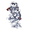 5e6yC  5e70C 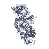 1m7xS S: Starting model for refinement C: citing same article ( |
|---|---|
| Similar structure data |


-Links
 PDBj
PDBj


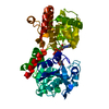
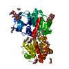
- Assembly
| Deposited unit | 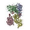
| ||||||||
|---|---|---|---|---|---|---|---|---|---|
| 1 | 
| ||||||||
| 2 | 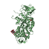
| ||||||||
| 3 | 
| ||||||||
| 4 | 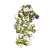
| ||||||||
| Unit cell |
|
-Components
| #1: Protein | Mass: 71277.633 Da / Num. of mol.: 4 Source method: isolated from a genetically manipulated source Source: (gene. exp.) Strain: E24377A / ETEC / Gene: glgB, EcE24377A_3911 / Production host: References: UniProt: A7ZSW5, 1,4-alpha-glucan branching enzyme #2: Polysaccharide | Cycloheptakis-(1-4)-(alpha-D-glucopyranose) / beta-cyclodextrin 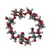  Source method: isolated from a genetically manipulated source Details: cyclic oligosaccharide / References: beta-cyclodextrin #3: Chemical | ChemComp-GOL /   Mass: 92.094 Da / Num. of mol.: 7 / Source method: obtained synthetically / Formula: C3H8O3 Mass: 92.094 Da / Num. of mol.: 7 / Source method: obtained synthetically / Formula: C3H8O3#4: Water | ChemComp-HOH / |  Mass: 18.015 Da / Num. of mol.: 1837 / Source method: isolated from a natural source / Formula: H2O Mass: 18.015 Da / Num. of mol.: 1837 / Source method: isolated from a natural source / Formula: H2O |
|---|
-Experimental details
-Experiment
| Experiment | Method: X-RAY DIFFRACTION |
|---|
- Sample preparation
| Crystal | Density Matthews: 3.22 Å3/Da / Density % sol: 61.82 % |
|---|---|
| Crystal grow | Temperature: 277 K / Method: vapor diffusion / pH: 7.2 / Details: 0.1 M NaHEPES, pH = 7.2 |
-Data collection
| Diffraction | Mean temperature: 100 K |
|---|---|
| Diffraction source | Source: SYNCHROTRON / Site: APS  / Beamline: 21-ID-D / Wavelength: 0.97876 Å / Beamline: 21-ID-D / Wavelength: 0.97876 Å |
| Detector | Type: MARMOSAIC 300 mm CCD / Detector: CCD / Date: Feb 15, 2008 |
| Radiation | Protocol: SINGLE WAVELENGTH / Monochromatic (M) / Laue (L): M / Scattering type: x-ray |
| Radiation wavelength | Wavelength: 0.97876 Å / Relative weight: 1 |
| Reflection | Resolution: 1.878→50 Å / % possible obs: 99.5 % / Redundancy: 4 % / Net I/σ(I): 25 |
- Processing
| Software |
| |||||||||||||||||||||||||||||||||||||||||||||||||||||||||||||||||||||||||||||||||||||||||||||||||||||||||||||||||||||||||||||||||||||||||||||||||||||||||||||||||||||||||||||||||||||||||||||||||||||||||||||||||||||||||
|---|---|---|---|---|---|---|---|---|---|---|---|---|---|---|---|---|---|---|---|---|---|---|---|---|---|---|---|---|---|---|---|---|---|---|---|---|---|---|---|---|---|---|---|---|---|---|---|---|---|---|---|---|---|---|---|---|---|---|---|---|---|---|---|---|---|---|---|---|---|---|---|---|---|---|---|---|---|---|---|---|---|---|---|---|---|---|---|---|---|---|---|---|---|---|---|---|---|---|---|---|---|---|---|---|---|---|---|---|---|---|---|---|---|---|---|---|---|---|---|---|---|---|---|---|---|---|---|---|---|---|---|---|---|---|---|---|---|---|---|---|---|---|---|---|---|---|---|---|---|---|---|---|---|---|---|---|---|---|---|---|---|---|---|---|---|---|---|---|---|---|---|---|---|---|---|---|---|---|---|---|---|---|---|---|---|---|---|---|---|---|---|---|---|---|---|---|---|---|---|---|---|---|---|---|---|---|---|---|---|---|---|---|---|---|---|---|---|---|
| Refinement | Method to determine structure: MOLECULAR REPLACEMENT Starting model: 1M7X Resolution: 1.878→44.927 Å / SU ML: 0.54 / Cross valid method: THROUGHOUT / σ(F): 1.34 / Phase error: 24.22 / Stereochemistry target values: ML
| |||||||||||||||||||||||||||||||||||||||||||||||||||||||||||||||||||||||||||||||||||||||||||||||||||||||||||||||||||||||||||||||||||||||||||||||||||||||||||||||||||||||||||||||||||||||||||||||||||||||||||||||||||||||||
| Solvent computation | Shrinkage radii: 0.73 Å / VDW probe radii: 1 Å / Solvent model: FLAT BULK SOLVENT MODEL / Bsol: 49.416 Å2 / ksol: 0.358 e/Å3 | |||||||||||||||||||||||||||||||||||||||||||||||||||||||||||||||||||||||||||||||||||||||||||||||||||||||||||||||||||||||||||||||||||||||||||||||||||||||||||||||||||||||||||||||||||||||||||||||||||||||||||||||||||||||||
| Displacement parameters |
| |||||||||||||||||||||||||||||||||||||||||||||||||||||||||||||||||||||||||||||||||||||||||||||||||||||||||||||||||||||||||||||||||||||||||||||||||||||||||||||||||||||||||||||||||||||||||||||||||||||||||||||||||||||||||
| Refinement step | Cycle: LAST / Resolution: 1.878→44.927 Å
| |||||||||||||||||||||||||||||||||||||||||||||||||||||||||||||||||||||||||||||||||||||||||||||||||||||||||||||||||||||||||||||||||||||||||||||||||||||||||||||||||||||||||||||||||||||||||||||||||||||||||||||||||||||||||
| Refine LS restraints |
| |||||||||||||||||||||||||||||||||||||||||||||||||||||||||||||||||||||||||||||||||||||||||||||||||||||||||||||||||||||||||||||||||||||||||||||||||||||||||||||||||||||||||||||||||||||||||||||||||||||||||||||||||||||||||
| LS refinement shell |
|
