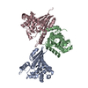
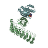
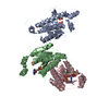
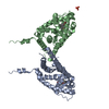
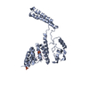
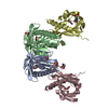
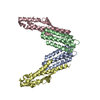
Entry Database : PDB / ID : 5e6pTitle PlexinB2 cytoplasmic region/PDZ-RhoGEF PDZ domain complex Plexin-B2 Rho guanine nucleotide exchange factor 11 Keywords / / / / Function / homology Function Domain/homology Component
/ / / / / / / / / / / / / / / / / / / / / / / / / / / / / / / / / / / / / / / / / / / / / / / / / / / / / / / / / / / / / / / / / / / / / / / / / / / / / / / / / / / / / / / / / / / / / / / / / / / / / / / / / / / / / / / / / / / / / / / / / / / / / / / Biological species Mus musculus (house mouse)Homo sapiens (human)Method / / / Resolution : 3.215 Å Authors Pascoe, H.G. / Zhang, X. Funding support Organization Grant number Country National Institutes of Health/National Institute of General Medical Sciences (NIH/NIGMS) GM088197 National Institutes of Health/National Institute of General Medical Sciences (NIH/NIGMS) GM031954 National Institutes of Health/National Institute of General Medical Sciences (NIH/NIGMS) GM008203
Journal : Proc.Natl.Acad.Sci.USA / Year : 2015Title : Secondary PDZ domain-binding site on class B plexins enhances the affinity for PDZ-RhoGEF.Authors : Pascoe, H.G. / Gutowski, S. / Chen, H. / Brautigam, C.A. / Chen, Z. / Sternweis, P.C. / Zhang, X. History Deposition Oct 10, 2015 Deposition site / Processing site Revision 1.0 Nov 18, 2015 Provider / Type Revision 1.1 Dec 16, 2015 Group Revision 1.2 Sep 27, 2017 Group / Database references / Derived calculationsCategory / pdbx_audit_support / pdbx_struct_oper_listItem / _pdbx_audit_support.funding_organization / _pdbx_struct_oper_list.symmetry_operationRevision 1.3 Dec 25, 2019 Group / Category / Item Revision 1.4 Sep 27, 2023 Group / Database references / Refinement descriptionCategory chem_comp_atom / chem_comp_bond ... chem_comp_atom / chem_comp_bond / database_2 / pdbx_initial_refinement_model Item / _database_2.pdbx_database_accession
Show all Show less
 Movie
Movie Controller


 Open data
 Basic information
 Components
 Keywords
 Function and homology information
 Homo sapiens (human)
 X-RAY DIFFRACTION /
  Authors
 United States, 3items
  Citation
 Structure visualization
 Downloads & links
 Other downloads
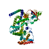


 PDBj
PDBj


 Assembly
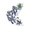

 Sample preparation
 Processing