 Movie
Movie Controller

[English] 日本語
 Yorodumi
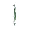
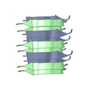
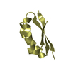
- PDB-5e5x: Structure of the amyloid forming peptide ANFLVH (residues 13-18) ... -
+ Open data

- Basic information
| Entry | Database: PDB / ID: 5e5x | ||||||
|---|---|---|---|---|---|---|---|
| Title | Structure of the amyloid forming peptide ANFLVH (residues 13-18) from islet amyloid polypeptide | ||||||
 Components Components | ANFLVH (residues 13-18) from islet amyloid polypeptide | ||||||
 Keywords Keywords | de novo protein / membrane protein / amyloid proto-fibril / Protein Fibril | ||||||
| Biological species |  Homo sapiens (human) Homo sapiens (human) | ||||||
| Method |  X-RAY DIFFRACTION / SYNCHROTRON / MOLECULAR REPLACEMENT / Resolution: 1.61 Å X-RAY DIFFRACTION / SYNCHROTRON / MOLECULAR REPLACEMENT / Resolution: 1.61 Å | ||||||
 Authors Authors | Soriaga, A.B. / Eisenberg, D. | ||||||
 Citation Citation | Journal: J.Phys.Chem.B / Year: 2016 Title: Crystal Structures of IAPP Amyloidogenic Segments Reveal a Novel Packing Motif of Out-of-Register Beta Sheets. Authors: Soriaga, A.B. / Sangwan, S. / Macdonald, R. / Sawaya, M.R. / Eisenberg, D. | ||||||
| History |
|
- Structure visualization
| Structure viewer | Molecule:  MolmilJmol/JSmol MolmilJmol/JSmol |
|---|
- Downloads & links
-Download
| PDBx/mmCIF format | 5e5x.cif.gz | 9.4 KB | Display | PDBx/mmCIF format |
|---|---|---|---|---|
| PDB format | pdb5e5x.ent.gz | 4.6 KB | Display | PDB format |
| PDBx/mmJSON format | 5e5x.json.gz | Tree view | PDBx/mmJSON format | |
| Others |  Other downloads Other downloads |
-Validation report
| Summary document | 5e5x_validation.pdf.gz | 401.2 KB | Display | wwPDB validaton report |
|---|---|---|---|---|
| Full document | 5e5x_full_validation.pdf.gz | 401.2 KB | Display | |
| Data in XML | 5e5x_validation.xml.gz | 2.3 KB | Display | |
| Data in CIF | 5e5x_validation.cif.gz | 2.3 KB | Display | |
| Arichive directory | https://data.pdbj.org/pub/pdb/validation_reports/e5/5e5xftp://data.pdbj.org/pub/pdb/validation_reports/e5/5e5x | HTTPS FTP |
-Related structure data


-Links
 PDBj
PDBj
- Assembly
| Deposited unit | 
| ||||||||
|---|---|---|---|---|---|---|---|---|---|
| 1 | x 10
| ||||||||
| Unit cell |
| ||||||||
| Details | BIOLOGICAL UNIT DISPLAYS ONLY A PORTION OF THE CRYSTAL LATTICE TO DEMONSTRATE THE CRYSTAL PACKING CONTENT. THE CRYSTAL PACKING IS FORMED BY A REPETITION IN BOTH DIRECTIONS OF THE PORTION INDICATED IN REMARK 350. |
-Components
| #1: Protein/peptide | Mass: 700.805 Da / Num. of mol.: 1 / Source method: obtained synthetically / Source: (synth.) Homo sapiens (human) |
|---|
-Experimental details
-Experiment
| Experiment | Method: X-RAY DIFFRACTION |
|---|
- Sample preparation
| Crystal | Density Matthews: 1.31 Å3/Da / Density % sol: 6.32 % |
|---|---|
| Crystal grow | Temperature: 291 K / Method: vapor diffusion, hanging drop Details: 20 mg/ml in water and mixed with 10% (w/v) PEG-8000, 0.1 M Na/K phosphate pH 6.2, and 0.2 M NaCl |
-Data collection
| Diffraction | Mean temperature: 291 K |
|---|---|
| Diffraction source | Source: SYNCHROTRON / Site: APS  / Beamline: 24-ID-E / Wavelength: 0.979 Å / Beamline: 24-ID-E / Wavelength: 0.979 Å |
| Detector | Type: ADSC QUANTUM 315 / Detector: CCD / Date: Nov 24, 2009 |
| Radiation | Protocol: SINGLE WAVELENGTH / Monochromatic (M) / Laue (L): M / Scattering type: x-ray |
| Radiation wavelength | Wavelength: 0.979 Å / Relative weight: 1 |
| Reflection | Resolution: 1.6→90 Å / Num. obs: 433 / % possible obs: 93.32 % / Redundancy: 3.1 % / Rmerge(I) obs: 0.147 / Net I/σ(I): 6.55 |
| Reflection shell | Resolution: 1.6→1.72 Å / Mean I/σ(I) obs: 9.7 / % possible all: 87 |
- Processing
| Software |
| ||||||||||||||||||||||||
|---|---|---|---|---|---|---|---|---|---|---|---|---|---|---|---|---|---|---|---|---|---|---|---|---|---|
| Refinement | Method to determine structure: MOLECULAR REPLACEMENT / Resolution: 1.61→19.863 Å / SU ML: 1.29 / Cross valid method: FREE R-VALUE / σ(F): 0.31 / Phase error: 17.45 / Stereochemistry target values: LS_WUNIT_K1
| ||||||||||||||||||||||||
| Solvent computation | Shrinkage radii: 0.9 Å / VDW probe radii: 1.11 Å / Solvent model: FLAT BULK SOLVENT MODEL / Bsol: 0 Å2 / ksol: 0 e/Å3 | ||||||||||||||||||||||||
| Displacement parameters |
| ||||||||||||||||||||||||
| Refinement step | Cycle: LAST / Resolution: 1.61→19.863 Å /
| ||||||||||||||||||||||||
| Refine LS restraints |
| ||||||||||||||||||||||||
| LS refinement shell | Resolution: 1.6101→19.8645 Å
|
