 Movie
Movie Controller


+ Open data

- Basic information
| Entry | Database: PDB / ID: 5e4v | |||||||||||||||||||||
|---|---|---|---|---|---|---|---|---|---|---|---|---|---|---|---|---|---|---|---|---|---|---|
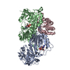
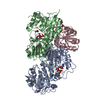
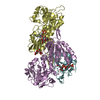
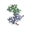
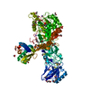
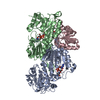
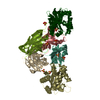
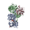
| Title | Crystal structure of measles N0-P complex | |||||||||||||||||||||
 Components Components | Nucleoprotein,Phosphoprotein | |||||||||||||||||||||
 Keywords Keywords | VIRAL PROTEIN / paramyxovirus / nucleocapsid protein / RNA-binding protein | |||||||||||||||||||||
| Function / homology |  Function and homology information Function and homology informationhost cell viral nucleoid / symbiont-mediated perturbation of host gene expression / positive regulation of viral transcription / helical viral capsid / Hsp70 protein binding / viral genome replication / molecular condensate scaffold activity / viral nucleocapsid / molecular adaptor activity / host cell cytoplasm ...host cell viral nucleoid / symbiont-mediated perturbation of host gene expression / positive regulation of viral transcription / helical viral capsid / Hsp70 protein binding / viral genome replication / molecular condensate scaffold activity / viral nucleocapsid / molecular adaptor activity / host cell cytoplasm / ribonucleoprotein complex / RNA-directed RNA polymerase activity / DNA-templated transcription / structural molecule activity / RNA binding Similarity search - Function | |||||||||||||||||||||
| Biological species |   Measles virus Measles virus | |||||||||||||||||||||
| Method |  X-RAY DIFFRACTION / SYNCHROTRON / MOLECULAR REPLACEMENT / Resolution: 2.71 Å X-RAY DIFFRACTION / SYNCHROTRON / MOLECULAR REPLACEMENT / Resolution: 2.71 Å | |||||||||||||||||||||
 Authors Authors | Guryanov, S.G. / Liljeroos, L. / Kasaragod, P. / Kajander, T. / Butcher, S.J. | |||||||||||||||||||||
| Funding support |  Finland, 6items Finland, 6items
| |||||||||||||||||||||
 Citation Citation | Journal: J.Virol. / Year: 2015 Title: Crystal Structure of the Measles Virus Nucleoprotein Core in Complex with an N-Terminal Region of Phosphoprotein. Authors: Guryanov, S.G. / Liljeroos, L. / Kasaragod, P. / Kajander, T. / Butcher, S.J. | |||||||||||||||||||||
| History |
|
- Structure visualization
| Structure viewer | Molecule: MolmilJmol/JSmol |
|---|
- Downloads & links
-Download
| PDBx/mmCIF format | 5e4v.cif.gz | 225.1 KB | Display | PDBx/mmCIF format |
|---|---|---|---|---|
| PDB format | pdb5e4v.ent.gz | 183.3 KB | Display | PDB format |
| PDBx/mmJSON format | 5e4v.json.gz | Tree view | PDBx/mmJSON format | |
| Others |  Other downloads Other downloads |
-Validation report
| Arichive directory | https://data.pdbj.org/pub/pdb/validation_reports/e4/5e4vftp://data.pdbj.org/pub/pdb/validation_reports/e4/5e4v | HTTPS FTP |
|---|
-Related structure data
| Related structure data |  4co6S S: Starting model for refinement |
|---|---|
| Similar structure data |
-Links
 PDBj
PDBj

- Assembly
| Deposited unit | 
| ||||||||
|---|---|---|---|---|---|---|---|---|---|
| 1 | 
| ||||||||
| Unit cell |
|
-Components
| #1: Protein | Mass: 48446.988 Da / Num. of mol.: 1 Fragment: nucleoprotein core domain with phosphoprotein binding peptide fused,nucleoprotein core domain with phosphoprotein binding peptide fused Source method: isolated from a genetically manipulated source Details: First residue remained from TEV cleavage site. Residues 2-389 correspond to measles nucleoprotein GenBank ID AAA18995.1 aa 21-408. Residues 390-437 correspond to measles phosphoprotein ...Details: First residue remained from TEV cleavage site. Residues 2-389 correspond to measles nucleoprotein GenBank ID AAA18995.1 aa 21-408. Residues 390-437 correspond to measles phosphoprotein GenBank ID AAF85668.1 aa 1-48 (although the full sequence of the protein in our virus strain (Halonen strain) differs by D512N variation compared to AAF85668.1, the difference does not affect the part of the sequence used in the experiment). Source: (gene. exp.) Measles virus (strain Edmonston B), (gene. exp.) Measles virus (strain Edmonston-Moraten vaccine)Strain: Edmonston B, Edmonston-Moraten vaccine / Gene: N, NP / Plasmid: pET22b / Production host:  |
|---|---|
| #2: Water | ChemComp-HOH /  Mass: 18.015 Da / Num. of mol.: 9 / Source method: isolated from a natural source / Formula: H2O Mass: 18.015 Da / Num. of mol.: 9 / Source method: isolated from a natural source / Formula: H2O |
-Experimental details
-Experiment
| Experiment | Method: X-RAY DIFFRACTION / Number of used crystals: 1 |
|---|
- Sample preparation
| Crystal | Density Matthews: 2.33 Å3/Da / Density % sol: 47.24 % |
|---|---|
| Crystal grow | Temperature: 295 K / Method: vapor diffusion, sitting drop / pH: 5.2 / Details: PEG 4000, sodium citrate |
-Data collection
| Diffraction | Mean temperature: 100 K | ||||||||||||||||||||||||
|---|---|---|---|---|---|---|---|---|---|---|---|---|---|---|---|---|---|---|---|---|---|---|---|---|---|
| Diffraction source | Source: SYNCHROTRON / Site: Diamond  / Beamline: I03 / Wavelength: 0.9763 Å / Beamline: I03 / Wavelength: 0.9763 Å | ||||||||||||||||||||||||
| Detector | Type: DECTRIS PILATUS 6M / Detector: PIXEL / Date: Jan 24, 2015 / Details: focused beam | ||||||||||||||||||||||||
| Radiation | Protocol: SINGLE WAVELENGTH / Monochromatic (M) / Laue (L): M / Scattering type: x-ray | ||||||||||||||||||||||||
| Radiation wavelength | Wavelength: 0.9763 Å / Relative weight: 1 | ||||||||||||||||||||||||
| Reflection | Resolution: 2.71→94.07 Å / Num. obs: 12618 / % possible obs: 100 % / Redundancy: 5.1 % / Biso Wilson estimate: 62.32 Å2 / CC1/2: 0.996 / Rmerge(I) obs: 0.123 / Rpim(I) all: 0.059 / Net I/σ(I): 7.5 / Num. measured all: 63741 / Scaling rejects: 2 | ||||||||||||||||||||||||
| Reflection shell | Diffraction-ID: 1 / Rejects: _ / % possible all: 100
|
- Processing
| Software |
| |||||||||||||||||||||||||||||||||||||||||||||||||||||||||||||||||||||||||||
|---|---|---|---|---|---|---|---|---|---|---|---|---|---|---|---|---|---|---|---|---|---|---|---|---|---|---|---|---|---|---|---|---|---|---|---|---|---|---|---|---|---|---|---|---|---|---|---|---|---|---|---|---|---|---|---|---|---|---|---|---|---|---|---|---|---|---|---|---|---|---|---|---|---|---|---|---|
| Refinement | Method to determine structure: MOLECULAR REPLACEMENT Starting model: 4CO6 Resolution: 2.71→78.93 Å / FOM work R set: 0.7854 / SU ML: 0.42 / Cross valid method: FREE R-VALUE / σ(F): 1.36 / Phase error: 27.65 / Stereochemistry target values: ML
| |||||||||||||||||||||||||||||||||||||||||||||||||||||||||||||||||||||||||||
| Solvent computation | Shrinkage radii: 1.01 Å / VDW probe radii: 1.1 Å / Solvent model: FLAT BULK SOLVENT MODEL / Bsol: 49.99 Å2 / ksol: 0.377 e/Å3 | |||||||||||||||||||||||||||||||||||||||||||||||||||||||||||||||||||||||||||
| Displacement parameters | Biso max: 259.59 Å2 / Biso mean: 68.86 Å2 / Biso min: 21.31 Å2
| |||||||||||||||||||||||||||||||||||||||||||||||||||||||||||||||||||||||||||
| Refinement step | Cycle: final / Resolution: 2.71→78.93 Å
| |||||||||||||||||||||||||||||||||||||||||||||||||||||||||||||||||||||||||||
| Refine LS restraints |
| |||||||||||||||||||||||||||||||||||||||||||||||||||||||||||||||||||||||||||
| LS refinement shell | Refine-ID: X-RAY DIFFRACTION / Total num. of bins used: 5 / % reflection obs: 100 %
| |||||||||||||||||||||||||||||||||||||||||||||||||||||||||||||||||||||||||||
| Refinement TLS params. | Method: refined / Refine-ID: X-RAY DIFFRACTION
| |||||||||||||||||||||||||||||||||||||||||||||||||||||||||||||||||||||||||||
| Refinement TLS group |
|
