 Movie
Movie Controller

[English] 日本語
 Yorodumi
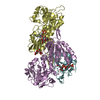
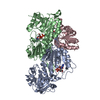
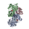
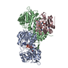
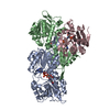
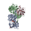
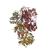
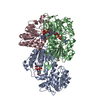
- PDB-1u28: R. rubrum transhydrogenase asymmetric complex (dI.NAD+)2(dIII.NADP+)1 -
+ Open data

- Basic information
| Entry | Database: PDB / ID: 1u28 | ||||||
|---|---|---|---|---|---|---|---|
| Title | R. rubrum transhydrogenase asymmetric complex (dI.NAD+)2(dIII.NADP+)1 | ||||||
 Components Components | (NAD(P) transhydrogenase subunit ...) x 2 | ||||||
 Keywords Keywords | OXIDOREDUCTASE / NAD(P) transhydrogenase subunits / NAD+ / NADP+ | ||||||
| Function / homology |  Function and homology information Function and homology informationNAD(P)+ transhydrogenase (Si-specific) activity / proton-translocating NAD(P)+ transhydrogenase activity / proton-translocating NAD(P)+ transhydrogenase / NADH binding / NADPH regeneration / NAD+ binding / membrane => GO:0016020 / NAD binding / NADP binding / protein dimerization activity / plasma membrane Similarity search - Function | ||||||
| Biological species |  Rhodospirillum rubrum (bacteria) Rhodospirillum rubrum (bacteria) | ||||||
| Method |  X-RAY DIFFRACTION / SYNCHROTRON / MOLECULAR REPLACEMENT / Resolution: 2.3 Å X-RAY DIFFRACTION / SYNCHROTRON / MOLECULAR REPLACEMENT / Resolution: 2.3 Å | ||||||
 Authors Authors | Mather, O.C. / Singh, A. / van Boxel, G.I. / White, S.A. / Jackson, J.B. | ||||||
 Citation Citation | Journal: Biochemistry / Year: 2004 Title: Active-site conformational changes associated with hydride transfer in proton-translocating transhydrogenase. Authors: Mather, O.C. / Singh, A. / van Boxel, G.I. / White, S.A. / Jackson, J.B. | ||||||
| History |
|
- Structure visualization
| Structure viewer | Molecule: MolmilJmol/JSmol |
|---|
- Downloads & links
-Download
| PDBx/mmCIF format | 1u28.cif.gz | 184.5 KB | Display | PDBx/mmCIF format |
|---|---|---|---|---|
| PDB format | pdb1u28.ent.gz | 146.3 KB | Display | PDB format |
| PDBx/mmJSON format | 1u28.json.gz | Tree view | PDBx/mmJSON format | |
| Others |  Other downloads Other downloads |
-Validation report
| Arichive directory | https://data.pdbj.org/pub/pdb/validation_reports/u2/1u28ftp://data.pdbj.org/pub/pdb/validation_reports/u2/1u28 | HTTPS FTP |
|---|
-Related structure data
| Related structure data |  1u2dC  1u2gC  1u31C  1hzzS S: Starting model for refinement C: citing same article ( |
|---|---|
| Similar structure data |


-Links
 PDBj
PDBj

- Assembly
| Deposited unit | 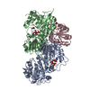
| ||||||||
|---|---|---|---|---|---|---|---|---|---|
| 1 |
| ||||||||
| Unit cell |
|
-Components
-NAD(P) transhydrogenase subunit ... , 2 types, 3 molecules ABC
| #1: Protein | Mass: 40324.785 Da / Num. of mol.: 2 Source method: isolated from a genetically manipulated source Source: (gene. exp.) Rhodospirillum rubrum (bacteria) / Gene: pntAA, nntA1 / Production host: References: UniProt: Q60164, UniProt: Q2RSB2*PLUS, EC: 1.6.1.2 #2: Protein | | Mass: 21485.510 Da / Num. of mol.: 1 / Fragment: Residues 262-464 Source method: isolated from a genetically manipulated source Source: (gene. exp.) Rhodospirillum rubrum (bacteria) / Gene: PNTB, nntb / Production host: References: UniProt: Q59765, UniProt: Q2RSB4*PLUS, EC: 1.6.1.2 |
|---|
-Non-polymers , 4 types, 115 molecules 


| #3: Chemical |  Mass: 663.425 Da / Num. of mol.: 2 / Source method: obtained synthetically / Formula: C21H27N7O14P2 / Comment: NAD*YM Mass: 663.425 Da / Num. of mol.: 2 / Source method: obtained synthetically / Formula: C21H27N7O14P2 / Comment: NAD*YM#4: Chemical |  Mass: 92.094 Da / Num. of mol.: 2 / Source method: obtained synthetically / Formula: C3H8O3 Mass: 92.094 Da / Num. of mol.: 2 / Source method: obtained synthetically / Formula: C3H8O3#5: Chemical | ChemComp-NAP / |  Mass: 743.405 Da / Num. of mol.: 1 / Source method: obtained synthetically / Formula: C21H28N7O17P3 Mass: 743.405 Da / Num. of mol.: 1 / Source method: obtained synthetically / Formula: C21H28N7O17P3#6: Water | ChemComp-HOH / | Mass: 18.015 Da / Num. of mol.: 110 / Source method: isolated from a natural source / Formula: H2O |
|---|
-Experimental details
-Experiment
| Experiment | Method: X-RAY DIFFRACTION / Number of used crystals: 1 |
|---|
- Sample preparation
| Crystal | Density Matthews: 2.65 Å3/Da / Density % sol: 53.56 % |
|---|---|
| Crystal grow | Temperature: 277 K / Method: vapor diffusion, sitting drop / pH: 6.5 Details: PEG-8K, AMMONIUM SULPHATE, pH 6.5, VAPOR DIFFUSION, SITTING DROP, temperature 277K |
-Data collection
| Diffraction | Mean temperature: 100 K |
|---|---|
| Diffraction source | Source: SYNCHROTRON / Site: ESRF  / Beamline: ID14-1 / Wavelength: 0.934 Å / Beamline: ID14-1 / Wavelength: 0.934 Å |
| Detector | Type: ADSC QUANTUM 4 / Detector: CCD |
| Radiation | Protocol: SINGLE WAVELENGTH / Monochromatic (M) / Laue (L): M / Scattering type: x-ray |
| Radiation wavelength | Wavelength: 0.934 Å / Relative weight: 1 |
| Reflection | Resolution: 2.3→49 Å / Num. obs: 47505 / % possible obs: 96.7 % / Redundancy: 4.5 % / Rsym value: 0.077 / Net I/σ(I): 11.6 |
- Processing
| Software |
| |||||||||||||||||||||
|---|---|---|---|---|---|---|---|---|---|---|---|---|---|---|---|---|---|---|---|---|---|---|
| Refinement | Method to determine structure: MOLECULAR REPLACEMENT Starting model: pdb entry 1HZZ Resolution: 2.3→49 Å / Rfactor Rfree: 0.275 / Rfactor Rwork: 0.241 / Isotropic thermal model: Isotropic / Cross valid method: THROUGHOUT | |||||||||||||||||||||
| Displacement parameters | Biso mean: 53.05 Å2 | |||||||||||||||||||||
| Refinement step | Cycle: LAST / Resolution: 2.3→49 Å
| |||||||||||||||||||||
| Refine LS restraints |
|
