Mass: 18.015 Da / Num. of mol.: 60 / Source method: isolated from a natural source / Formula: D2O
-
Experimental details
-
Experiment
Experiment
Method
Number of used crystals
X-RAY DIFFRACTION
1
NEUTRON DIFFRACTION
1
-
Sample preparation
Crystal
Density Matthews: 2.3 Å3/Da / Density % sol: 46.59 %
Crystal grow
Temperature: 293 K / Method: vapor diffusion, sitting drop / pH: 5.5 Details: Large crystals were grown in 9-well glass plates using Hampton sandwich box set-up.
-
Data collection
Diffraction
ID
Mean temperature (K)
Crystal-ID
2
293
1
1
100
1
Diffraction source
Source
Site
Beamline
ID
Wavelength (Å)
NUCLEAR REACTOR
FRM II
BIODIFF
2
2.66
SYNCHROTRON
MAX II
I911-2
1
1.04
Detector
Type
ID
Detector
Date
FUJI
2
IMAGE PLATE
Nov 25, 2014
MAR CCD 165 mm
1
CCD
Oct 28, 2014
Radiation
ID
Protocol
Monochromatic (M) / Laue (L)
Scattering type
Wavelength-ID
2
SINGLEWAVELENGTH
M
neutron
2
1
SINGLEWAVELENGTH
M
x-ray
1
Radiation wavelength
ID
Wavelength (Å)
Relative weight
1
2.66
1
2
1.04
1
Reflection
Entry-ID: 5DPN
Resolution (Å)
Num. obs
% possible obs (%)
Redundancy (%)
Rmerge(I) obs
Diffraction-ID
Net I/σ(I)
1.6-45
20530
91.1
3
0.041
2
6.6
1.6-29.3
21610
97.7
5.5
0.093
1
28.6
Reflection shell
Resolution (Å)
Redundancy (%)
Rmerge(I) obs
Mean I/σ(I) obs
Num. unique all
Diffraction-ID
% possible all
1.6-1.7
5.5
0.231
7.8
3389
1
97.2
1.6-1.65
2.5
0.449
1.8
1485
2
97.2
-
Processing
Software
Name
Version
Classification
PHENIX
1.9_1692
refinement
DENZO
datareduction
SCALEPACK
datascaling
Coot
modelbuilding
Refinement
SU ML: 0.19 / Cross valid method: THROUGHOUT / Method to determine structure: MOLECULAR REPLACEMENT / Shrinkage radii: 0.9 Å / VDW probe radii: 1.11 Å / Starting model: 4bj0
In the structure databanks used in Yorodumi, some data are registered as the other names, "COVID-19 virus" and "2019-nCoV". Here are the details of the virus and the list of structure data.
Jan 31, 2019. EMDB accession codes are about to change! (news from PDBe EMDB page)
EMDB accession codes are about to change! (news from PDBe EMDB page)
The allocation of 4 digits for EMDB accession codes will soon come to an end. Whilst these codes will remain in use, new EMDB accession codes will include an additional digit and will expand incrementally as the available range of codes is exhausted. The current 4-digit format prefixed with “EMD-” (i.e. EMD-XXXX) will advance to a 5-digit format (i.e. EMD-XXXXX), and so on. It is currently estimated that the 4-digit codes will be depleted around Spring 2019, at which point the 5-digit format will come into force.
The EM Navigator/Yorodumi systems omit the EMD- prefix.
Related info.:Q: What is EMD? / ID/Accession-code notation in Yorodumi/EM Navigator
Yorodumi is a browser for structure data from EMDB, PDB, SASBDB, etc.
This page is also the successor to EM Navigator detail page, and also detail information page/front-end page for Omokage search.
The word "yorodu" (or yorozu) is an old Japanese word meaning "ten thousand". "mi" (miru) is to see.
Related info.:EMDB / PDB / SASBDB / Comparison of 3 databanks / Yorodumi Search / Aug 31, 2016. New EM Navigator & Yorodumi / Yorodumi Papers / Jmol/JSmol / Function and homology information / Changes in new EM Navigator and Yorodumi
 Movie
Movie Controller

 Yorodumi
 Open data
 Basic information
 Components
 Keywords
 Function and homology information
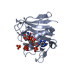
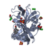
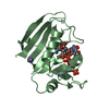
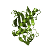
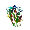
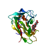
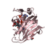
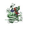
 Rhodothermus marinus (bacteria)
 X-RAY DIFFRACTION / NEUTRON DIFFRACTION / NUCLEAR REACTOR /
  Authors
 Citation
 Structure visualization
 Downloads & links
 Other downloads


 PDBj
PDBj


 Assembly
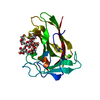

 Mass: 40.078 Da / Num. of mol.: 1 / Source method: obtained synthetically / Formula: Ca
 Mass: 18.015 Da / Num. of mol.: 60 / Source method: isolated from a natural source / Formula: D2O
 Sample preparation

 Processing