 Movie
Movie Controller

[English] 日本語
 Yorodumi
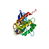
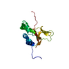
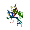
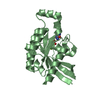
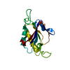
- PDB-5dmp: Structure of the Archaeal NHEJ Phosphoesterase from Methanocella ... -
+ Open data

- Basic information
| Entry | Database: PDB / ID: 5dmp | ||||||
|---|---|---|---|---|---|---|---|
| Title | Structure of the Archaeal NHEJ Phosphoesterase from Methanocella paludicola. | ||||||
 Components Components | Uncharacterized protein | ||||||
 Keywords Keywords | HYDROLASE / Archaeal Proteins / Biocatalysis / DNA Repair Enzymes / Phosphoric Diester Hydrolases / Phosphoric Monoester Hydrolases | ||||||
| Function / homology | DNA ligase D, 3'-phosphoesterase domain / DNA Ligase D 3'-phosphoesterase domain / oxido(dioxo)vanadium / DNA ligase D 3'-phosphoesterase domain-containing protein Function and homology information Function and homology information | ||||||
| Biological species |  Methanocella paludicola (archaea) Methanocella paludicola (archaea) | ||||||
| Method |  X-RAY DIFFRACTION / SYNCHROTRON / MOLECULAR REPLACEMENT / molecular replacement / Resolution: 1.793 Å X-RAY DIFFRACTION / SYNCHROTRON / MOLECULAR REPLACEMENT / molecular replacement / Resolution: 1.793 Å | ||||||
 Authors Authors | Brissett, N.C. / Bartlett, E.J. / Doherty, A.J. | ||||||
| Funding support |  United Kingdom, 1items United Kingdom, 1items
| ||||||
 Citation Citation | Journal: Nucleic Acids Res. / Year: 2016 Title: Molecular basis for DNA strand displacement by NHEJ repair polymerases. Authors: Bartlett, E.J. / Brissett, N.C. / Plocinski, P. / Carlberg, T. / Doherty, A.J. | ||||||
| History |
|
- Structure visualization
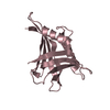
| Structure viewer | Molecule: MolmilJmol/JSmol |
|---|
- Downloads & links
-Download
| PDBx/mmCIF format | 5dmp.cif.gz | 52.6 KB | Display | PDBx/mmCIF format |
|---|---|---|---|---|
| PDB format | pdb5dmp.ent.gz | 35.9 KB | Display | PDB format |
| PDBx/mmJSON format | 5dmp.json.gz | Tree view | PDBx/mmJSON format | |
| Others |  Other downloads Other downloads |
-Validation report
| Arichive directory | https://data.pdbj.org/pub/pdb/validation_reports/dm/5dmpftp://data.pdbj.org/pub/pdb/validation_reports/dm/5dmp | HTTPS FTP |
|---|
-Related structure data
| Related structure data |  5dmuC  3n9bS S: Starting model for refinement C: citing same article ( |
|---|---|
| Similar structure data |


-Links
 PDBj
PDBj- Assembly
| Deposited unit | 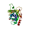
| ||||||||
|---|---|---|---|---|---|---|---|---|---|
| 1 |
| ||||||||
| Unit cell |
|
-Components
| #1: Protein | Mass: 19062.793 Da / Num. of mol.: 1 / Fragment: UNP residues 30-198 Source method: isolated from a genetically manipulated source Source: (gene. exp.) Methanocella paludicola (archaea) / Gene: MCP_2127 / Plasmid: pET28 / Production host:  | ||||||
|---|---|---|---|---|---|---|---|
| #2: Chemical | ChemComp-EDO /   Mass: 62.068 Da / Num. of mol.: 7 / Source method: obtained synthetically / Formula: C2H6O2 Mass: 62.068 Da / Num. of mol.: 7 / Source method: obtained synthetically / Formula: C2H6O2#3: Chemical | ChemComp-MG / |   Mass: 24.305 Da / Num. of mol.: 1 / Source method: obtained synthetically / Formula: Mg Mass: 24.305 Da / Num. of mol.: 1 / Source method: obtained synthetically / Formula: Mg#4: Chemical | ChemComp-VN4 / | 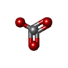  Mass: 98.940 Da / Num. of mol.: 1 / Source method: obtained synthetically / Formula: VO3 Mass: 98.940 Da / Num. of mol.: 1 / Source method: obtained synthetically / Formula: VO3#5: Water | ChemComp-HOH / |  Mass: 18.015 Da / Num. of mol.: 90 / Source method: isolated from a natural source / Formula: H2O Mass: 18.015 Da / Num. of mol.: 90 / Source method: isolated from a natural source / Formula: H2O |
-Experimental details
-Experiment
| Experiment | Method: X-RAY DIFFRACTION / Number of used crystals: 1 |
|---|
- Sample preparation
| Crystal | Density Matthews: 2.59 Å3/Da / Density % sol: 52.52 % |
|---|---|
| Crystal grow | Temperature: 285 K / Method: vapor diffusion, sitting drop / pH: 7.5 / Details: 200 mM magnesium sulfate, 20% (w/v) PEG 3350 |
-Data collection
| Diffraction | Mean temperature: 100 K | ||||||||||||||||||||||||||||||||||||||||||||||||||||||||||||||||||||||||||||||||||||||||||||||||||||||||||||||
|---|---|---|---|---|---|---|---|---|---|---|---|---|---|---|---|---|---|---|---|---|---|---|---|---|---|---|---|---|---|---|---|---|---|---|---|---|---|---|---|---|---|---|---|---|---|---|---|---|---|---|---|---|---|---|---|---|---|---|---|---|---|---|---|---|---|---|---|---|---|---|---|---|---|---|---|---|---|---|---|---|---|---|---|---|---|---|---|---|---|---|---|---|---|---|---|---|---|---|---|---|---|---|---|---|---|---|---|---|---|---|---|
| Diffraction source | Source: SYNCHROTRON / Site: Diamond / Beamline: I03 / Wavelength: 0.9763 Å | ||||||||||||||||||||||||||||||||||||||||||||||||||||||||||||||||||||||||||||||||||||||||||||||||||||||||||||||
| Detector | Type: DECTRIS PILATUS 300K / Detector: PIXEL / Date: May 27, 2011 | ||||||||||||||||||||||||||||||||||||||||||||||||||||||||||||||||||||||||||||||||||||||||||||||||||||||||||||||
| Radiation | Protocol: SINGLE WAVELENGTH / Monochromatic (M) / Laue (L): M / Scattering type: x-ray | ||||||||||||||||||||||||||||||||||||||||||||||||||||||||||||||||||||||||||||||||||||||||||||||||||||||||||||||
| Radiation wavelength | Wavelength: 0.9763 Å / Relative weight: 1 | ||||||||||||||||||||||||||||||||||||||||||||||||||||||||||||||||||||||||||||||||||||||||||||||||||||||||||||||
| Reflection | Resolution: 1.793→49.424 Å / Num. all: 18976 / Num. obs: 18976 / % possible obs: 98.9 % / Redundancy: 5 % / Rpim(I) all: 0.025 / Rrim(I) all: 0.056 / Rsym value: 0.05 / Net I/av σ(I): 8.17 / Net I/σ(I): 16.4 / Num. measured all: 95174 | ||||||||||||||||||||||||||||||||||||||||||||||||||||||||||||||||||||||||||||||||||||||||||||||||||||||||||||||
| Reflection shell | Diffraction-ID: 1 / Rejects: _
|
-Phasing
| Phasing | Method: molecular replacement | |||||||||
|---|---|---|---|---|---|---|---|---|---|---|
| Phasing MR | Rfactor: 32.66 / Model details: Phaser MODE: MR_AUTO
|
- Processing
| Software |
| ||||||||||||||||||||||||||||||||||||||||||||||||||||||||||||
|---|---|---|---|---|---|---|---|---|---|---|---|---|---|---|---|---|---|---|---|---|---|---|---|---|---|---|---|---|---|---|---|---|---|---|---|---|---|---|---|---|---|---|---|---|---|---|---|---|---|---|---|---|---|---|---|---|---|---|---|---|---|
| Refinement | Method to determine structure: MOLECULAR REPLACEMENT Starting model: 3n9b Resolution: 1.793→49.42 Å / Cor.coef. Fo:Fc: 0.968 / Cor.coef. Fo:Fc free: 0.961 / WRfactor Rfree: 0.215 / WRfactor Rwork: 0.1838 / FOM work R set: 0.8475 / SU B: 2.571 / SU ML: 0.079 / SU R Cruickshank DPI: 0.1092 / SU Rfree: 0.1057 / Cross valid method: THROUGHOUT / σ(F): 0 / ESU R: 0.109 / ESU R Free: 0.106 / Stereochemistry target values: MAXIMUM LIKELIHOOD Details: HYDROGENS HAVE BEEN USED IF PRESENT IN THE INPUT U VALUES : REFINED INDIVIDUALLY
| ||||||||||||||||||||||||||||||||||||||||||||||||||||||||||||
| Solvent computation | Ion probe radii: 0.8 Å / Shrinkage radii: 0.8 Å / VDW probe radii: 1.2 Å / Solvent model: BABINET MODEL WITH MASK | ||||||||||||||||||||||||||||||||||||||||||||||||||||||||||||
| Displacement parameters | Biso max: 87.83 Å2 / Biso mean: 37.484 Å2 / Biso min: 21.38 Å2
| ||||||||||||||||||||||||||||||||||||||||||||||||||||||||||||
| Refinement step | Cycle: final / Resolution: 1.793→49.42 Å
| ||||||||||||||||||||||||||||||||||||||||||||||||||||||||||||
| Refine LS restraints |
| ||||||||||||||||||||||||||||||||||||||||||||||||||||||||||||
| LS refinement shell | Resolution: 1.793→1.84 Å / Total num. of bins used: 20
|
