[English] 日本語
Yorodumi
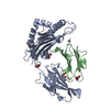
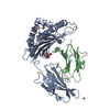
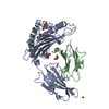
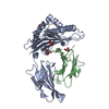
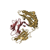
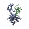
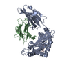
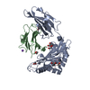
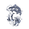
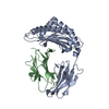
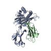
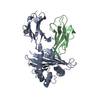
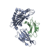
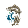
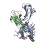
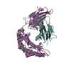
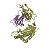
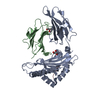
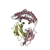
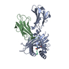
- PDB-5deg: Crystal structure of B*27:06 bound to the pVIPR peptide -

+
Open data


ID or keywords:

Loading...

-
Basic information

Entry
Database: PDB / ID: 5deg
TitleCrystal structure of B*27:06 bound to the pVIPR peptide
Components
  • Beta-2-microglobulin
  • MHC class I antigen
  • Peptide derived of VASOACTIVE INTESTINAL POLYPEPTIDE RECEPTOR 1 (pVIPR)
KeywordsIMMUNE SYSTEM / IMMUNE SYSTEM-COMPLEX / MHC MAJOR HISTOCOMPATIBILITY COMPLEX) / HLA- B*2706
Function / homology
Function and homology information


vasoactive intestinal polypeptide receptor activity / G protein-coupled peptide receptor activity / regulation of interleukin-12 production / regulation of dendritic cell differentiation / regulation of T cell anergy / regulation of interleukin-6 production / antigen processing and presentation / peptide hormone binding / G protein-coupled receptor signaling pathway, coupled to cyclic nucleotide second messenger / TAP binding ...vasoactive intestinal polypeptide receptor activity / G protein-coupled peptide receptor activity / regulation of interleukin-12 production / regulation of dendritic cell differentiation / regulation of T cell anergy / regulation of interleukin-6 production / antigen processing and presentation / peptide hormone binding / G protein-coupled receptor signaling pathway, coupled to cyclic nucleotide second messenger / TAP binding / protection from natural killer cell mediated cytotoxicity / detection of bacterium / secretory granule membrane / positive regulation of ferrous iron binding / positive regulation of transferrin receptor binding / positive regulation of receptor binding / early endosome lumen / Nef mediated downregulation of MHC class I complex cell surface expression / DAP12 interactions / negative regulation of receptor binding / antigen processing and presentation of endogenous peptide antigen via MHC class I via ER pathway, TAP-independent / antigen processing and presentation of endogenous peptide antigen via MHC class Ib / lumenal side of endoplasmic reticulum membrane / cellular response to iron ion / Endosomal/Vacuolar pathway / Antigen Presentation: Folding, assembly and peptide loading of class I MHC / cellular response to iron(III) ion / antigen processing and presentation of exogenous protein antigen via MHC class Ib, TAP-dependent / negative regulation of forebrain neuron differentiation / regulation of erythrocyte differentiation / adenylate cyclase-modulating G protein-coupled receptor signaling pathway / ER to Golgi transport vesicle membrane / peptide antigen assembly with MHC class I protein complex / regulation of iron ion transport / response to molecule of bacterial origin / MHC class I peptide loading complex / HFE-transferrin receptor complex / T cell mediated cytotoxicity / antigen processing and presentation of endogenous peptide antigen via MHC class I / positive regulation of T cell cytokine production / MHC class I protein complex / defense response / multicellular organismal-level iron ion homeostasis / Glucagon-type ligand receptors / negative regulation of neurogenesis / peptide antigen assembly with MHC class II protein complex / positive regulation of receptor-mediated endocytosis / MHC class II protein complex / cellular response to nicotine / positive regulation of T cell mediated cytotoxicity / specific granule lumen / recycling endosome membrane / phagocytic vesicle membrane / positive regulation of cellular senescence / peptide antigen binding / negative regulation of epithelial cell proliferation / antigen processing and presentation of exogenous peptide antigen via MHC class II / Immunoregulatory interactions between a Lymphoid and a non-Lymphoid cell / Interferon gamma signaling / positive regulation of immune response / Modulation by Mtb of host immune system / Interferon alpha/beta signaling / sensory perception of smell / positive regulation of T cell activation / positive regulation of protein binding / tertiary granule lumen / DAP12 signaling / negative regulation of neuron projection development / MHC class II protein complex binding / late endosome membrane / iron ion transport / ER-Phagosome pathway / early endosome membrane / protein-folding chaperone binding / T cell differentiation in thymus / protein refolding / G alpha (s) signalling events / protein homotetramerization / intracellular iron ion homeostasis / adaptive immune response / amyloid fibril formation / membrane => GO:0016020 / learning or memory / cell surface receptor signaling pathway / receptor complex / immune response / G protein-coupled receptor signaling pathway / Amyloid fiber formation / endoplasmic reticulum lumen / lysosomal membrane / external side of plasma membrane / Golgi membrane / innate immune response / focal adhesion / signaling receptor binding / positive regulation of cell population proliferation / Neutrophil degranulation / SARS-CoV-2 activates/modulates innate and adaptive immune responses / structural molecule activity / Golgi apparatus
Similarity search - Function
GPCR, family 2, vasoactive intestinal peptide receptor 1 / GPCR, family 2, vasoactive intestinal peptide receptor / G-protein coupled receptors family 2 signature 1. / Hormone receptor domain / GPCR, family 2, extracellular hormone receptor domain / G-protein coupled receptors family 2 profile 1. / Domain present in hormone receptors / GPCR family 2, extracellular hormone receptor domain superfamily / G-protein coupled receptors family 2 signature 2. / GPCR, family 2, secretin-like, conserved site ...GPCR, family 2, vasoactive intestinal peptide receptor 1 / GPCR, family 2, vasoactive intestinal peptide receptor / G-protein coupled receptors family 2 signature 1. / Hormone receptor domain / GPCR, family 2, extracellular hormone receptor domain / G-protein coupled receptors family 2 profile 1. / Domain present in hormone receptors / GPCR family 2, extracellular hormone receptor domain superfamily / G-protein coupled receptors family 2 signature 2. / GPCR, family 2, secretin-like, conserved site / GPCR, family 2, secretin-like / 7 transmembrane receptor (Secretin family) / GPCR, family 2-like / G-protein coupled receptors family 2 profile 2. / MHC class I, alpha chain, C-terminal / MHC_I C-terminus / MHC class I-like antigen recognition-like / Murine Class I Major Histocompatibility Complex, H2-DB; Chain A, domain 1 / MHC class I alpha chain, alpha1 alpha2 domains / Class I Histocompatibility antigen, domains alpha 1 and 2 / Beta-2-Microglobulin / MHC class I-like antigen recognition-like / MHC class I-like antigen recognition-like superfamily / MHC classes I/II-like antigen recognition protein / Immunoglobulin/major histocompatibility complex, conserved site / Immunoglobulins and major histocompatibility complex proteins signature. / Immunoglobulin C-Type / Immunoglobulin C1-set / Immunoglobulin C1-set domain / Ig-like domain profile. / Immunoglobulin-like domain / Immunoglobulin-like domain superfamily / Immunoglobulins / Immunoglobulin-like fold / Immunoglobulin-like / Sandwich / 2-Layer Sandwich / Mainly Beta / Alpha Beta
Similarity search - Domain/homology
HLA class I histocompatibility antigen, B alpha chain / Vasoactive intestinal polypeptide receptor 1 / Beta-2-microglobulin / HLA-B alpha chain (B*2706)
Similarity search - Component
Biological speciesHomo sapiens (human)
MethodX-RAY DIFFRACTION / SYNCHROTRON / MOLECULAR REPLACEMENT / Resolution: 1.83 Å
AuthorsLoll, B. / Fabian, H. / Hee, C.S. / Huser, H. / Uchanska-Ziegler, B. / Ziegler, A.
Funding support Germany, 2items
OrganizationGrant numberCountry
German Research FoundationSFB 449 Germany
German Research FoundationUC 8/1-3 Germany
CitationJournal: Arthritis Rheumatol / Year: 2016
Title: Increased Conformational Flexibility of HLA-B*27 Subtypes Associated With Ankylosing Spondylitis.
Authors: Loll, B. / Fabian, H. / Huser, H. / Hee, C.S. / Ziegler, A. / Uchanska-Ziegler, B. / Ziegler, A.
History
DepositionAug 25, 2015Deposition site: RCSB / Processing site: PDBE
Revision 1.0Nov 18, 2015Provider: repository / Type: Initial release
Revision 1.1Jan 20, 2016Group: Database references
Revision 1.2Jan 27, 2016Group: Database references
Revision 1.3Dec 18, 2019Group: Author supporting evidence / Database references
Category: citation / pdbx_audit_support / pdbx_related_exp_data_set
Item: _citation.country / _pdbx_audit_support.funding_organization
Revision 1.4Jan 10, 2024Group: Data collection / Database references / Refinement description
Category: chem_comp_atom / chem_comp_bond ...chem_comp_atom / chem_comp_bond / database_2 / pdbx_initial_refinement_model
Item: _database_2.pdbx_DOI / _database_2.pdbx_database_accession

-
Structure visualization

Structure viewerMolecule:
MolmilJmol/JSmol

Downloads & links

-
Assembly

Deposited unit
A: MHC class I antigen
B: Beta-2-microglobulin
C: Peptide derived of VASOACTIVE INTESTINAL POLYPEPTIDE RECEPTOR 1 (pVIPR)
hetero molecules


Theoretical massNumber of molelcules
Total (without water)45,4045
Polymers45,2203
Non-polymers1842
Water7,062392
1


  • Idetical with deposited unit
  • defined by software
TypeNameSymmetry operationNumber
identity operation1_555x,y,z1
Buried area5300 Å2
ΔGint-16 kcal/mol
Surface area19140 Å2
MethodPISA
Unit cell
Length a, b, c (Å)50.768, 81.684, 64.433
Angle α, β, γ (deg.)90.00, 107.53, 90.00
Int Tables number4
Space group name H-MP1211

-
Components

#1: Protein MHC class I antigen


Mass: 31941.133 Da / Num. of mol.: 1 / Fragment: residues 25-300
Source method: isolated from a genetically manipulated source
Source: (gene. exp.) Homo sapiens (human) / Gene: HLA-B / Plasmid: PHN1 / Production host: Escherichia coli BL21 (bacteria) / References: UniProt: Q7YQB0, UniProt: P01889*PLUS
#2: Protein Beta-2-microglobulin


Mass: 11879.356 Da / Num. of mol.: 1
Source method: isolated from a genetically manipulated source
Source: (gene. exp.) Homo sapiens (human) / Gene: B2M, CDABP0092, HDCMA22P / Plasmid: PHN1 / Production host: Escherichia coli BL21 (bacteria) / References: UniProt: P61769
#3: Protein/peptide Peptide derived of VASOACTIVE INTESTINAL POLYPEPTIDE RECEPTOR 1 (pVIPR)


Mass: 1399.694 Da / Num. of mol.: 1 / Source method: obtained synthetically / Source: (synth.) Homo sapiens (human) / References: UniProt: P32241*PLUS
#4: Chemical ChemComp-GOL / GLYCEROL / GLYCERIN / PROPANE-1,2,3-TRIOL


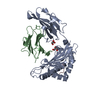
Mass: 92.094 Da / Num. of mol.: 2 / Source method: obtained synthetically / Formula: C3H8O3
#5: Water ChemComp-HOH / water


Mass: 18.015 Da / Num. of mol.: 392 / Source method: isolated from a natural source / Formula: H2O

-
Experimental details

-
Experiment

ExperimentMethod: X-RAY DIFFRACTION

-
Sample preparation

CrystalDensity Matthews: 2.9 Å3/Da / Density % sol: 57.5 %
Crystal growTemperature: 291 K / Method: vapor diffusion, hanging drop / pH: 7 / Details: 20%(w/v) PEG 8000, 100 mM Tris-HCl pH 7.0 / Temp details: 291

-
Data collection

DiffractionMean temperature: 100 K
Diffraction sourceSource: SYNCHROTRON / Site: MPG/DESY, HAMBURG / Beamline: BW6 / Wavelength: 1.005 Å
DetectorType: MARRESEARCH / Detector: CCD / Date: May 10, 2005
RadiationProtocol: SINGLE WAVELENGTH / Monochromatic (M) / Laue (L): M / Scattering type: x-ray
Radiation wavelengthWavelength: 1.005 Å / Relative weight: 1
ReflectionResolution: 1.83→30 Å / Num. obs: 43847 / % possible obs: 99.1 % / Redundancy: 3.6 % / Biso Wilson estimate: 20.4 Å2 / Rsym value: 0.036 / Net I/σ(I): 34.7
Reflection shellResolution: 1.83→1.9 Å / Redundancy: 3.4 % / Mean I/σ(I) obs: 6 / % possible all: 92.2

-
Processing

Software
NameVersionClassification
REFMAC5.5.0109refinement
DENZOdata reduction
SCALEPACKdata scaling
PHASERphasing
RefinementMethod to determine structure: MOLECULAR REPLACEMENT
Starting model: 2a83
Resolution: 1.83→20 Å / Cor.coef. Fo:Fc: 0.957 / Cor.coef. Fo:Fc free: 0.945 / SU B: 4.015 / SU ML: 0.066 / Cross valid method: THROUGHOUT / ESU R: 0.123 / ESU R Free: 0.11 / Stereochemistry target values: MAXIMUM LIKELIHOOD / Details: HYDROGENS HAVE BEEN ADDED IN THE RIDING POSITIONS
RfactorNum. reflection% reflectionSelection details
Rfree0.19308 2205 5 %RANDOM
Rwork0.17097 ---
obs0.17214 41622 99.57 %-
Solvent computationIon probe radii: 0.8 Å / Shrinkage radii: 0.8 Å / VDW probe radii: 1.4 Å / Solvent model: MASK
Displacement parametersBiso mean: 19.591 Å2
Baniso -1Baniso -2Baniso -3
1-0.29 Å20 Å20.48 Å2
2---0.77 Å20 Å2
3---0.76 Å2
Refinement stepCycle: 1 / Resolution: 1.83→20 Å
ProteinNucleic acidLigandSolventTotal
Num. atoms3190 0 12 392 3594
Refine LS restraints
Refine-IDTypeDev idealDev ideal targetNumber
X-RAY DIFFRACTIONr_bond_refined_d0.0110.0213588
X-RAY DIFFRACTIONr_bond_other_d0.0010.022569
X-RAY DIFFRACTIONr_angle_refined_deg1.2671.9324897
X-RAY DIFFRACTIONr_angle_other_deg0.79836189
X-RAY DIFFRACTIONr_dihedral_angle_1_deg5.9855423
X-RAY DIFFRACTIONr_dihedral_angle_2_deg29.37222.938211
X-RAY DIFFRACTIONr_dihedral_angle_3_deg12.99215628
X-RAY DIFFRACTIONr_dihedral_angle_4_deg16.9491543
X-RAY DIFFRACTIONr_chiral_restr0.0820.2498
X-RAY DIFFRACTIONr_gen_planes_refined0.0050.0214014
X-RAY DIFFRACTIONr_gen_planes_other0.0010.02802
X-RAY DIFFRACTIONr_nbd_refined
X-RAY DIFFRACTIONr_nbd_other
X-RAY DIFFRACTIONr_nbtor_refined
X-RAY DIFFRACTIONr_nbtor_other
X-RAY DIFFRACTIONr_xyhbond_nbd_refined
X-RAY DIFFRACTIONr_xyhbond_nbd_other
X-RAY DIFFRACTIONr_metal_ion_refined
X-RAY DIFFRACTIONr_metal_ion_other
X-RAY DIFFRACTIONr_symmetry_vdw_refined
X-RAY DIFFRACTIONr_symmetry_vdw_other
X-RAY DIFFRACTIONr_symmetry_hbond_refined
X-RAY DIFFRACTIONr_symmetry_hbond_other
X-RAY DIFFRACTIONr_symmetry_metal_ion_refined
X-RAY DIFFRACTIONr_symmetry_metal_ion_other
X-RAY DIFFRACTIONr_mcbond_it0.6081.52034
X-RAY DIFFRACTIONr_mcbond_other0.161.5800
X-RAY DIFFRACTIONr_mcangle_it1.10523338
X-RAY DIFFRACTIONr_mcangle_other
X-RAY DIFFRACTIONr_scbond_it1.65931554
X-RAY DIFFRACTIONr_scbond_other
X-RAY DIFFRACTIONr_scangle_it2.7274.51543
X-RAY DIFFRACTIONr_scangle_other
X-RAY DIFFRACTIONr_long_range_B_refined
X-RAY DIFFRACTIONr_long_range_B_other
X-RAY DIFFRACTIONr_rigid_bond_restr
X-RAY DIFFRACTIONr_sphericity_free
X-RAY DIFFRACTIONr_sphericity_bonded
LS refinement shellResolution: 1.833→1.88 Å / Total num. of bins used: 20
RfactorNum. reflection% reflection
Rfree0.29 143 -
Rwork0.271 2989 -
obs--96.46 %
Refinement TLS params.

Method: refined / Refine-ID: X-RAY DIFFRACTION

IDL112)L122)L132)L222)L232)L332)S11 (Å °)S12 (Å °)S13 (Å °)S21 (Å °)S22 (Å °)S23 (Å °)S31 (Å °)S32 (Å °)S33 (Å °)T112)T122)T132)T222)T232)T332)Origin x (Å)Origin y (Å)Origin z (Å)
12.2713-0.1148-0.34660.78940.03050.55250.0282-0.1637-0.00950.1071-0.00840.01570.0039-0.023-0.01980.0243-0.00620.00320.01690.00220.001510.39017.806233.7922
21.10010.29420.28023.36343.50548.3457-0.00310.3224-0.0551-0.5877-0.06310.0838-0.1689-0.11430.06620.15980.0103-0.00620.1256-0.01160.0537-9.8373.58274.8955
34.0162-1.13261.19752.9952-1.12762.10270.01210.31340.3556-0.1783-0.03470.1102-0.0368-0.05280.02260.0693-0.0061-0.00560.06050.0440.05520.618621.918313.5989
428.0544-0.9311-8.75070.03350.28892.7305-0.2131-2.6162-0.31680.01510.0935-0.00460.06780.81920.11960.0948-0.0008-0.00610.27920.04610.109215.32337.175341.2273
Refinement TLS group
IDRefine-IDRefine TLS-IDAuth asym-IDAuth seq-ID
1X-RAY DIFFRACTION1A1 - 185
2X-RAY DIFFRACTION2A186 - 276
3X-RAY DIFFRACTION3B0 - 99
4X-RAY DIFFRACTION4C1 - 9

+
About Yorodumi

-
News

-
Feb 9, 2022. New format data for meta-information of EMDB entries

New format data for meta-information of EMDB entries

  • Version 3 of the EMDB header file is now the official format.
  • The previous official version 1.9 will be removed from the archive.

Related info.:EMDB header

External links:wwPDB to switch to version 3 of the EMDB data model

-
Aug 12, 2020. Covid-19 info

Covid-19 info

URL: https://pdbjlvh1.pdbj.org/emnavi/covid19.php

New page: Covid-19 featured information page in EM Navigator.

Related info.:Covid-19 info / Mar 5, 2020. Novel coronavirus structure data

+
Mar 5, 2020. Novel coronavirus structure data

Novel coronavirus structure data

Related info.:Yorodumi Speices / Aug 12, 2020. Covid-19 info

External links:COVID-19 featured content - PDBj / Molecule of the Month (242):Coronavirus Proteases

+
Jan 31, 2019. EMDB accession codes are about to change! (news from PDBe EMDB page)

EMDB accession codes are about to change! (news from PDBe EMDB page)

  • The allocation of 4 digits for EMDB accession codes will soon come to an end. Whilst these codes will remain in use, new EMDB accession codes will include an additional digit and will expand incrementally as the available range of codes is exhausted. The current 4-digit format prefixed with “EMD-” (i.e. EMD-XXXX) will advance to a 5-digit format (i.e. EMD-XXXXX), and so on. It is currently estimated that the 4-digit codes will be depleted around Spring 2019, at which point the 5-digit format will come into force.
  • The EM Navigator/Yorodumi systems omit the EMD- prefix.

Related info.:Q: What is EMD? / ID/Accession-code notation in Yorodumi/EM Navigator

External links:EMDB Accession Codes are Changing Soon! / Contact to PDBj

+
Jul 12, 2017. Major update of PDB

Major update of PDB

  • wwPDB released updated PDB data conforming to the new PDBx/mmCIF dictionary.
  • This is a major update changing the version number from 4 to 5, and with Remediation, in which all the entries are updated.
  • In this update, many items about electron microscopy experimental information are reorganized (e.g. em_software).
  • Now, EM Navigator and Yorodumi are based on the updated data.

External links:wwPDB Remediation / Enriched Model Files Conforming to OneDep Data Standards Now Available in the PDB FTP Archive

-
Yorodumi

Thousand views of thousand structures

  • Yorodumi is a browser for structure data from EMDB, PDB, SASBDB, etc.
  • This page is also the successor to EM Navigator detail page, and also detail information page/front-end page for Omokage search.
  • The word "yorodu" (or yorozu) is an old Japanese word meaning "ten thousand". "mi" (miru) is to see.

Related info.:EMDB / PDB / SASBDB / Comparison of 3 databanks / Yorodumi Search / Aug 31, 2016. New EM Navigator & Yorodumi / Yorodumi Papers / Jmol/JSmol / Function and homology information / Changes in new EM Navigator and Yorodumi

Read more