 Movie
Movie Controller

[English] 日本語
 Yorodumi
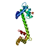
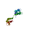
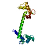
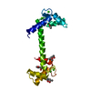
- PDB-5dbr: Ca2+ CaM with human cardiac Na+ channel (NaV1.5) inactivation gate -
+ Open data

- Basic information
| Entry | Database: PDB / ID: 5dbr | |||||||||||||||
|---|---|---|---|---|---|---|---|---|---|---|---|---|---|---|---|---|
| Title | Ca2+ CaM with human cardiac Na+ channel (NaV1.5) inactivation gate | |||||||||||||||
 Components Components |
| |||||||||||||||
 Keywords Keywords | Metal Binding Protein/Transport Protein / Calmodulin / NaV1.5 / Inactivation gate / Metal Binding Protein-Transport Protein complex | |||||||||||||||
| Function / homology |  Function and homology information Function and homology informationvoltage-gated sodium channel activity involved in AV node cell action potential / voltage-gated sodium channel activity involved in bundle of His cell action potential / voltage-gated sodium channel activity involved in Purkinje myocyte action potential / voltage-gated sodium channel activity involved in SA node cell action potential / bundle of His cell action potential / regulation of ventricular cardiac muscle cell membrane depolarization / AV node cell action potential / SA node cell action potential / AV node cell to bundle of His cell communication / membrane depolarization during SA node cell action potential ...voltage-gated sodium channel activity involved in AV node cell action potential / voltage-gated sodium channel activity involved in bundle of His cell action potential / voltage-gated sodium channel activity involved in Purkinje myocyte action potential / voltage-gated sodium channel activity involved in SA node cell action potential / bundle of His cell action potential / regulation of ventricular cardiac muscle cell membrane depolarization / AV node cell action potential / SA node cell action potential / AV node cell to bundle of His cell communication / membrane depolarization during SA node cell action potential / response to denervation involved in regulation of muscle adaptation / membrane depolarization during atrial cardiac muscle cell action potential / voltage-gated sodium channel activity involved in cardiac muscle cell action potential / regulation of atrial cardiac muscle cell membrane repolarization / cardiac ventricle development / brainstem development / : / : / : / regulation of atrial cardiac muscle cell membrane depolarization / : / positive regulation of action potential / atrial cardiac muscle cell action potential / positive regulation of protein autophosphorylation / : / membrane depolarization during AV node cell action potential / cardiac conduction system development / membrane depolarization during bundle of His cell action potential / telencephalon development / membrane depolarization during Purkinje myocyte cell action potential / membrane depolarization during cardiac muscle cell action potential / negative regulation of peptidyl-threonine phosphorylation / positive regulation of sodium ion transport / membrane depolarization during action potential / regulation of sodium ion transmembrane transport / : / ventricular cardiac muscle cell action potential / type 3 metabotropic glutamate receptor binding / regulation of ventricular cardiac muscle cell membrane repolarization / cardiac muscle cell action potential involved in contraction / voltage-gated sodium channel complex / regulation of cardiac muscle cell contraction / positive regulation of peptidyl-threonine phosphorylation / positive regulation of DNA binding / CaM pathway / Cam-PDE 1 activation / voltage-gated sodium channel activity / Sodium/Calcium exchangers / Interaction between L1 and Ankyrins / ankyrin binding / Calmodulin induced events / Reduction of cytosolic Ca++ levels / Activation of Ca-permeable Kainate Receptor / CREB1 phosphorylation through the activation of CaMKII/CaMKK/CaMKIV cascasde / Loss of phosphorylation of MECP2 at T308 / CREB1 phosphorylation through the activation of Adenylate Cyclase / negative regulation of high voltage-gated calcium channel activity / PKA activation / CaMK IV-mediated phosphorylation of CREB / Glycogen breakdown (glycogenolysis) / CLEC7A (Dectin-1) induces NFAT activation / response to corticosterone / negative regulation of ryanodine-sensitive calcium-release channel activity / organelle localization by membrane tethering / Activation of RAC1 downstream of NMDARs / : / autophagosome membrane docking / odontogenesis of dentin-containing tooth / regulation of synaptic vesicle exocytosis / negative regulation of calcium ion export across plasma membrane / regulation of ryanodine-sensitive calcium-release channel activity / regulation of cardiac muscle cell action potential / presynaptic endocytosis / sodium ion transport / Synthesis of IP3 and IP4 in the cytosol / positive regulation of protein serine/threonine kinase activity / Phase 0 - rapid depolarisation / Negative regulation of NMDA receptor-mediated neuronal transmission / Unblocking of NMDA receptors, glutamate binding and activation / RHO GTPases activate PAKs / calcineurin-mediated signaling / regulation of heart rate by cardiac conduction / nitric-oxide synthase binding / regulation of cell communication by electrical coupling involved in cardiac conduction / fibroblast growth factor binding / Ion transport by P-type ATPases / adenylate cyclase binding / Uptake and function of anthrax toxins / protein phosphatase activator activity / intercalated disc / Long-term potentiation / lateral plasma membrane / Calcineurin activates NFAT / Regulation of MECP2 expression and activity / DARPP-32 events / Smooth Muscle Contraction / regulation of synaptic vesicle endocytosis / detection of calcium ion / membrane depolarization / regulation of cardiac muscle contraction Similarity search - Function | |||||||||||||||
| Biological species |  Homo sapiens (human) Homo sapiens (human) | |||||||||||||||
| Method |  X-RAY DIFFRACTION / MOLECULAR REPLACEMENT / Resolution: 2.25 Å X-RAY DIFFRACTION / MOLECULAR REPLACEMENT / Resolution: 2.25 Å | |||||||||||||||
 Authors Authors | Johnson, C.N. / Thompson, M.K. / Chazin, W.J. | |||||||||||||||
| Funding support |  United States, 4items United States, 4items
| |||||||||||||||
 Citation Citation | Journal: To Be Published Title: Enhanced Understanding of Ca2+ Modulation of the Human Cardiac Sodium Channel: Tight Binding of Calmodulin to the Inactivation Gate Authors: Johnson, C.N. / Thompson, M.K. / Voehler, M. / Chazin, W.J. | |||||||||||||||
| History |
|
- Structure visualization
| Structure viewer | Molecule: MolmilJmol/JSmol |
|---|
- Downloads & links
-Download
| PDBx/mmCIF format | 5dbr.cif.gz | 48.3 KB | Display | PDBx/mmCIF format |
|---|---|---|---|---|
| PDB format | pdb5dbr.ent.gz | 31.4 KB | Display | PDB format |
| PDBx/mmJSON format | 5dbr.json.gz | Tree view | PDBx/mmJSON format | |
| Others |  Other downloads Other downloads |
-Validation report
| Arichive directory | https://data.pdbj.org/pub/pdb/validation_reports/db/5dbrftp://data.pdbj.org/pub/pdb/validation_reports/db/5dbr | HTTPS FTP |
|---|
-Related structure data
| Related structure data | 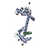 4djcS S: Starting model for refinement |
|---|---|
| Similar structure data |


-Links
 PDBj
PDBj


- Assembly
| Deposited unit | 
| ||||||||
|---|---|---|---|---|---|---|---|---|---|
| 1 |
| ||||||||
| Unit cell |
|
-Components
| #1: Protein | Mass: 16407.053 Da / Num. of mol.: 1 Source method: isolated from a genetically manipulated source Source: (gene. exp.) Homo sapiens (human)Gene: CALM1, CALM, CAM, CAM1, CALM2, CAM2, CAMB, CALM3, CALML2, CAM3, CAMC, CAMIII Production host:  | ||
|---|---|---|---|
| #2: Protein | Mass: 5898.891 Da / Num. of mol.: 1 / Fragment: UNP residues 1483-1529 Source method: isolated from a genetically manipulated source Source: (gene. exp.) Homo sapiens (human) / Gene: SCN5A / Production host: | ||
| #3: Chemical | ChemComp-CA /   Mass: 40.078 Da / Num. of mol.: 4 / Source method: obtained synthetically / Formula: Ca Mass: 40.078 Da / Num. of mol.: 4 / Source method: obtained synthetically / Formula: Ca#4: Water | ChemComp-HOH / |  Mass: 18.015 Da / Num. of mol.: 12 / Source method: isolated from a natural source / Formula: H2O Mass: 18.015 Da / Num. of mol.: 12 / Source method: isolated from a natural source / Formula: H2O |
-Experimental details
-Experiment
| Experiment | Method: X-RAY DIFFRACTION / Number of used crystals: 1 |
|---|
- Sample preparation
| Crystal | Density Matthews: 2.71 Å3/Da / Density % sol: 54.7 % |
|---|---|
| Crystal grow | Temperature: 298 K / Method: vapor diffusion, hanging drop / pH: 4 / Details: 0.2M sodium malonate pH 4.0, 20% w/v PEG 3350 |
-Data collection
| Diffraction | Mean temperature: 100 K |
|---|---|
| Diffraction source | Source: ROTATING ANODE / Type: BRUKER AXS MICROSTAR / Wavelength: 1.5418 Å |
| Detector | Type: Bruker Platinum 135 / Detector: CCD / Date: Jul 31, 2013 |
| Radiation | Protocol: SINGLE WAVELENGTH / Monochromatic (M) / Laue (L): M / Scattering type: x-ray |
| Radiation wavelength | Wavelength: 1.5418 Å / Relative weight: 1 |
| Reflection | Resolution: 2.25→50.3 Å / Num. obs: 9401 / % possible obs: 99.58 % / Redundancy: 15.6 % / Rmerge(I) obs: 0.0905 / Net I/σ(I): 18.4 |
| Reflection shell | Resolution: 2.25→2.3 Å / Redundancy: 10.56 % / Rmerge(I) obs: 0.4455 / Mean I/σ(I) obs: 4.1 / % possible all: 100 |
- Processing
| Software |
| |||||||||||||||||||||||||||||||||||||||||||||||||||||||||||||||||||||||||||
|---|---|---|---|---|---|---|---|---|---|---|---|---|---|---|---|---|---|---|---|---|---|---|---|---|---|---|---|---|---|---|---|---|---|---|---|---|---|---|---|---|---|---|---|---|---|---|---|---|---|---|---|---|---|---|---|---|---|---|---|---|---|---|---|---|---|---|---|---|---|---|---|---|---|---|---|---|
| Refinement | Method to determine structure: MOLECULAR REPLACEMENT Starting model: 4DJC Resolution: 2.25→50.3 Å / Cor.coef. Fo:Fc: 0.943 / Cor.coef. Fo:Fc free: 0.9 / SU B: 7.137 / SU ML: 0.174 / Cross valid method: THROUGHOUT / σ(F): 0 / ESU R: 0.261 / ESU R Free: 0.234 / Stereochemistry target values: MAXIMUM LIKELIHOOD Details: HYDROGENS HAVE BEEN ADDED IN THE RIDING POSITIONS U VALUES : REFINED INDIVIDUALLY
| |||||||||||||||||||||||||||||||||||||||||||||||||||||||||||||||||||||||||||
| Solvent computation | Ion probe radii: 0.8 Å / Shrinkage radii: 0.8 Å / VDW probe radii: 1.2 Å / Solvent model: MASK | |||||||||||||||||||||||||||||||||||||||||||||||||||||||||||||||||||||||||||
| Displacement parameters | Biso max: 82.68 Å2 / Biso mean: 39.957 Å2 / Biso min: 20 Å2
| |||||||||||||||||||||||||||||||||||||||||||||||||||||||||||||||||||||||||||
| Refinement step | Cycle: final / Resolution: 2.25→50.3 Å
| |||||||||||||||||||||||||||||||||||||||||||||||||||||||||||||||||||||||||||
| Refine LS restraints |
| |||||||||||||||||||||||||||||||||||||||||||||||||||||||||||||||||||||||||||
| LS refinement shell | Resolution: 2.25→2.308 Å / Total num. of bins used: 20
|
