 Movie
Movie Controller

[English] 日本語
 Yorodumi
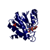
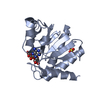
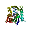
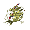
- PDB-5cuw: Crystal structure of Sortase E1 from Streptomyces coelicolor with... -
+ Open data

- Basic information
| Entry | Database: PDB / ID: 5cuw | ||||||
|---|---|---|---|---|---|---|---|
| Title | Crystal structure of Sortase E1 from Streptomyces coelicolor with tripeptide in the active site | ||||||
 Components Components | SrtE1 | ||||||
 Keywords Keywords | MEMBRANE PROTEIN / sortase / class E / SrtE / transpeptidase / aminoacyltransferase / cysteine endopeptidase / bacterial protein / protein binding / catalytic domain / beta barrel | ||||||
| Function / homology |  Function and homology information Function and homology information | ||||||
| Biological species |  Streptomyces coelicolor (bacteria) Streptomyces coelicolor (bacteria) | ||||||
| Method |  X-RAY DIFFRACTION / SYNCHROTRON / MOLECULAR REPLACEMENT / molecular replacement / Resolution: 1.887 Å X-RAY DIFFRACTION / SYNCHROTRON / MOLECULAR REPLACEMENT / molecular replacement / Resolution: 1.887 Å | ||||||
 Authors Authors | Kattke, M.D. / Cascio, D. / Sawaya, M.R. / Clubb, R.T. | ||||||
 Citation Citation | Journal: To be published Title: Crystal structure of the first class E sortase transpeptidase, SrtE1 from Streptomyces coelicolor Authors: Kattke, M.D. / Cascio, D. / Sawaya, M.R. / Elliot, M.A. / Clubb, R.T. | ||||||
| History |
|
- Structure visualization
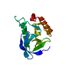
| Structure viewer | Molecule: MolmilJmol/JSmol |
|---|
- Downloads & links
-Download
| PDBx/mmCIF format | 5cuw.cif.gz | 79.1 KB | Display | PDBx/mmCIF format |
|---|---|---|---|---|
| PDB format | pdb5cuw.ent.gz | 57 KB | Display | PDB format |
| PDBx/mmJSON format | 5cuw.json.gz | Tree view | PDBx/mmJSON format | |
| Others |  Other downloads Other downloads |
-Validation report
| Arichive directory | https://data.pdbj.org/pub/pdb/validation_reports/cu/5cuwftp://data.pdbj.org/pub/pdb/validation_reports/cu/5cuw | HTTPS FTP |
|---|
-Related structure data
| Similar structure data |
|---|


-Links
 PDBj
PDBj- Assembly
| Deposited unit | 
| ||||||||
|---|---|---|---|---|---|---|---|---|---|
| 1 |
| ||||||||
| Unit cell |
| ||||||||
| Components on special symmetry positions |
|
-Components
| #1: Protein | Mass: 21249.221 Da / Num. of mol.: 1 / Fragment: UNP residues 162-352 Source method: isolated from a genetically manipulated source Source: (gene. exp.) Streptomyces coelicolor (strain ATCC BAA-471 / A3(2) / M145) (bacteria)Strain: ATCC BAA-471 / A3(2) / M145 / Gene: SCO3850 / Plasmid: pET-15b / Production host: |
|---|---|
| #2: Chemical | ChemComp-GOL /   Mass: 92.094 Da / Num. of mol.: 1 / Fragment: GOL / Source method: obtained synthetically / Formula: C3H8O3 Mass: 92.094 Da / Num. of mol.: 1 / Fragment: GOL / Source method: obtained synthetically / Formula: C3H8O3 |
| #3: Water | ChemComp-HOH /  Mass: 18.015 Da / Num. of mol.: 37 / Source method: isolated from a natural source / Formula: H2O Mass: 18.015 Da / Num. of mol.: 37 / Source method: isolated from a natural source / Formula: H2O |
-Experimental details
-Experiment
| Experiment | Method: X-RAY DIFFRACTION / Number of used crystals: 1 |
|---|
- Sample preparation
| Crystal | Density Matthews: 2.57 Å3/Da / Density % sol: 52.23 % |
|---|---|
| Crystal grow | Temperature: 277 K / Method: vapor diffusion, hanging drop / pH: 7.5 / Details: 50 mM HEPES, pH 7.5, 1.4 M sodium citrate |
-Data collection
| Diffraction | Mean temperature: 100 K | ||||||||||||||||||||||||||||||||||||||||||||||||||||||||||||||||||||||||||||||||||||||||||||||||||||||||||||||||||||||||||||||||||||||||||||||||||||||||||||||||||||||||||||||||||||||||||||||||||||||||||||||||||
|---|---|---|---|---|---|---|---|---|---|---|---|---|---|---|---|---|---|---|---|---|---|---|---|---|---|---|---|---|---|---|---|---|---|---|---|---|---|---|---|---|---|---|---|---|---|---|---|---|---|---|---|---|---|---|---|---|---|---|---|---|---|---|---|---|---|---|---|---|---|---|---|---|---|---|---|---|---|---|---|---|---|---|---|---|---|---|---|---|---|---|---|---|---|---|---|---|---|---|---|---|---|---|---|---|---|---|---|---|---|---|---|---|---|---|---|---|---|---|---|---|---|---|---|---|---|---|---|---|---|---|---|---|---|---|---|---|---|---|---|---|---|---|---|---|---|---|---|---|---|---|---|---|---|---|---|---|---|---|---|---|---|---|---|---|---|---|---|---|---|---|---|---|---|---|---|---|---|---|---|---|---|---|---|---|---|---|---|---|---|---|---|---|---|---|---|---|---|---|---|---|---|---|---|---|---|---|---|---|---|---|---|
| Diffraction source | Source: SYNCHROTRON / Site: APS  / Beamline: 24-ID-C / Wavelength: 0.9791 Å / Beamline: 24-ID-C / Wavelength: 0.9791 Å | ||||||||||||||||||||||||||||||||||||||||||||||||||||||||||||||||||||||||||||||||||||||||||||||||||||||||||||||||||||||||||||||||||||||||||||||||||||||||||||||||||||||||||||||||||||||||||||||||||||||||||||||||||
| Detector | Type: DECTRIS PILATUS 6M / Detector: PIXEL / Date: Oct 12, 2012 | ||||||||||||||||||||||||||||||||||||||||||||||||||||||||||||||||||||||||||||||||||||||||||||||||||||||||||||||||||||||||||||||||||||||||||||||||||||||||||||||||||||||||||||||||||||||||||||||||||||||||||||||||||
| Radiation | Protocol: SINGLE WAVELENGTH / Monochromatic (M) / Laue (L): M / Scattering type: x-ray | ||||||||||||||||||||||||||||||||||||||||||||||||||||||||||||||||||||||||||||||||||||||||||||||||||||||||||||||||||||||||||||||||||||||||||||||||||||||||||||||||||||||||||||||||||||||||||||||||||||||||||||||||||
| Radiation wavelength | Wavelength: 0.9791 Å / Relative weight: 1 | ||||||||||||||||||||||||||||||||||||||||||||||||||||||||||||||||||||||||||||||||||||||||||||||||||||||||||||||||||||||||||||||||||||||||||||||||||||||||||||||||||||||||||||||||||||||||||||||||||||||||||||||||||
| Reflection | Resolution: 1.88→43.53 Å / Num. obs: 17717 / % possible obs: 97.3 % / Observed criterion σ(I): -3 / Redundancy: 5.9 % / Biso Wilson estimate: 26.16 Å2 / Rmerge F obs: 0.987 / Rmerge(I) obs: 0.189 / Rrim(I) all: 0.207 / Χ2: 1.059 / Net I/σ(I): 6.82 / Num. measured all: 104280 | ||||||||||||||||||||||||||||||||||||||||||||||||||||||||||||||||||||||||||||||||||||||||||||||||||||||||||||||||||||||||||||||||||||||||||||||||||||||||||||||||||||||||||||||||||||||||||||||||||||||||||||||||||
| Reflection shell | Diffraction-ID: 1 / Rejects: _
|
-Phasing
| Phasing | Method: molecular replacement | |||||||||
|---|---|---|---|---|---|---|---|---|---|---|
| Phasing MR | Model details: Phaser MODE: MR_AUTO
|
- Processing
| Software |
| |||||||||||||||||||||||||||||||||||||||||||||||||
|---|---|---|---|---|---|---|---|---|---|---|---|---|---|---|---|---|---|---|---|---|---|---|---|---|---|---|---|---|---|---|---|---|---|---|---|---|---|---|---|---|---|---|---|---|---|---|---|---|---|---|
| Refinement | Method to determine structure: MOLECULAR REPLACEMENT / Resolution: 1.887→43.526 Å / SU ML: 0.25 / Cross valid method: FREE R-VALUE / σ(F): 1.32 / Phase error: 26.84 / Stereochemistry target values: ML
| |||||||||||||||||||||||||||||||||||||||||||||||||
| Solvent computation | Shrinkage radii: 0.9 Å / VDW probe radii: 1.11 Å / Solvent model: FLAT BULK SOLVENT MODEL | |||||||||||||||||||||||||||||||||||||||||||||||||
| Displacement parameters | Biso max: 108.74 Å2 / Biso mean: 33.7971 Å2 / Biso min: 15.57 Å2 | |||||||||||||||||||||||||||||||||||||||||||||||||
| Refinement step | Cycle: final / Resolution: 1.887→43.526 Å
| |||||||||||||||||||||||||||||||||||||||||||||||||
| Refine LS restraints |
| |||||||||||||||||||||||||||||||||||||||||||||||||
| LS refinement shell | Refine-ID: X-RAY DIFFRACTION / Total num. of bins used: 6
| |||||||||||||||||||||||||||||||||||||||||||||||||
| Refinement TLS params. | Method: refined / Origin x: -6.6234 Å / Origin y: -20.9557 Å / Origin z: 6.0701 Å
| |||||||||||||||||||||||||||||||||||||||||||||||||
| Refinement TLS group |
|
