 Movie
Movie Controller

[English] 日本語
 Yorodumi
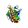
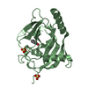
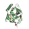
- PDB-5cph: Crystal structure of the ATP binding domain of S. aureus GyrB com... -
+ Open data

- Basic information
| Entry | Database: PDB / ID: 5cph | ||||||
|---|---|---|---|---|---|---|---|
| Title | Crystal structure of the ATP binding domain of S. aureus GyrB complexed with a fragment | ||||||
 Components Components | DNA gyrase subunit B | ||||||
 Keywords Keywords | ISOMERASE/ISOMERASE INHIBITOR / DNA gyrase / GyrB / fragment-based screening / structure-based design / ISOMERASE-ISOMERASE INHIBITOR complex | ||||||
| Function / homology |  Function and homology information Function and homology informationDNA negative supercoiling activity / DNA topoisomerase (ATP-hydrolysing) / DNA topological change / DNA-templated DNA replication / chromosome / response to antibiotic / DNA binding / ATP binding / metal ion binding / cytoplasm Similarity search - Function | ||||||
| Biological species |   Staphylococcus aureus (bacteria) Staphylococcus aureus (bacteria) | ||||||
| Method |  X-RAY DIFFRACTION / SYNCHROTRON / MOLECULAR REPLACEMENT / molecular replacement / Resolution: 1.2 Å X-RAY DIFFRACTION / SYNCHROTRON / MOLECULAR REPLACEMENT / molecular replacement / Resolution: 1.2 Å | ||||||
 Authors Authors | Andersen, O.A. / Barker, J. / Cheng, R.K. / Kahmann, J. / Felicetti, B. / Wood, M. / Scheich, C. / Mesleh, M. / Cross, J.B. / Zhang, J. ...Andersen, O.A. / Barker, J. / Cheng, R.K. / Kahmann, J. / Felicetti, B. / Wood, M. / Scheich, C. / Mesleh, M. / Cross, J.B. / Zhang, J. / Yang, Q. / Lippa, B. / Ryan, M.D. | ||||||
 Citation Citation | Journal: Bioorg.Med.Chem.Lett. / Year: 2016 Title: Fragment-based discovery of DNA gyrase inhibitors targeting the ATPase subunit of GyrB. Authors: Mesleh, M.F. / Cross, J.B. / Zhang, J. / Kahmann, J. / Andersen, O.A. / Barker, J. / Cheng, R.K. / Felicetti, B. / Wood, M. / Hadfield, A.T. / Scheich, C. / Moy, T.I. / Yang, Q. / Shotwell, ...Authors: Mesleh, M.F. / Cross, J.B. / Zhang, J. / Kahmann, J. / Andersen, O.A. / Barker, J. / Cheng, R.K. / Felicetti, B. / Wood, M. / Hadfield, A.T. / Scheich, C. / Moy, T.I. / Yang, Q. / Shotwell, J. / Nguyen, K. / Lippa, B. / Dolle, R. / Ryan, M.D. | ||||||
| History |
|
- Structure visualization
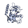
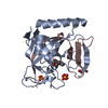
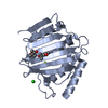
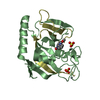
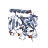
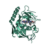
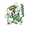
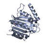
| Structure viewer | Molecule: MolmilJmol/JSmol |
|---|
- Downloads & links
-Download
| PDBx/mmCIF format | 5cph.cif.gz | 107.3 KB | Display | PDBx/mmCIF format |
|---|---|---|---|---|
| PDB format | pdb5cph.ent.gz | 80.2 KB | Display | PDB format |
| PDBx/mmJSON format | 5cph.json.gz | Tree view | PDBx/mmJSON format | |
| Others |  Other downloads Other downloads |
-Validation report
| Arichive directory | https://data.pdbj.org/pub/pdb/validation_reports/cp/5cphftp://data.pdbj.org/pub/pdb/validation_reports/cp/5cph | HTTPS FTP |
|---|
-Related structure data
| Related structure data |  5ctuC 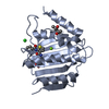 5ctwC  5ctxC  5ctyC 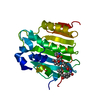 1kznS C: citing same article ( S: Starting model for refinement |
|---|---|
| Similar structure data |


-Links
 PDBj
PDBj


- Assembly
| Deposited unit | 
| ||||||||
|---|---|---|---|---|---|---|---|---|---|
| 1 | 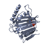
| ||||||||
| 2 | 
| ||||||||
| Unit cell |
| ||||||||
| Components on special symmetry positions |
|
-Components
| #1: Protein | Mass: 23931.602 Da / Num. of mol.: 2 Fragment: ATP binding domain, UNP residues 2-234 (delta 105-127) Source method: isolated from a genetically manipulated source Source: (gene. exp.) Staphylococcus aureus (bacteria) / Gene: gyrB / Plasmid: pET24 / Production host: #2: Chemical |   Mass: 222.242 Da / Num. of mol.: 2 / Source method: obtained synthetically / Formula: C14H10N2O Mass: 222.242 Da / Num. of mol.: 2 / Source method: obtained synthetically / Formula: C14H10N2O#3: Chemical |   Mass: 96.063 Da / Num. of mol.: 2 / Source method: obtained synthetically / Formula: SO4 Mass: 96.063 Da / Num. of mol.: 2 / Source method: obtained synthetically / Formula: SO4#4: Chemical | ChemComp-MPD / ( |   Mass: 118.174 Da / Num. of mol.: 1 / Source method: obtained synthetically / Formula: C6H14O2 / Comment: precipitant*YM Mass: 118.174 Da / Num. of mol.: 1 / Source method: obtained synthetically / Formula: C6H14O2 / Comment: precipitant*YM#5: Water | ChemComp-HOH / |  Mass: 18.015 Da / Num. of mol.: 522 / Source method: isolated from a natural source / Formula: H2O Mass: 18.015 Da / Num. of mol.: 522 / Source method: isolated from a natural source / Formula: H2O |
|---|
-Experimental details
-Experiment
| Experiment | Method: X-RAY DIFFRACTION / Number of used crystals: 1 |
|---|
- Sample preparation
| Crystal | Density Matthews: 2.11 Å3/Da / Density % sol: 41.58 % |
|---|---|
| Crystal grow | Temperature: 296 K / Method: vapor diffusion, hanging drop / pH: 7.6 Details: 40-43% MPD_P1K_P3350, 100 mM Mops/Na-Hepes, 100 mM Divalents |
-Data collection
| Diffraction | Mean temperature: 100 K | |||||||||||||||||||||||||||||||||||||||||||||||||||||||||||||||||||||||||||||||||||||||||||||||||||
|---|---|---|---|---|---|---|---|---|---|---|---|---|---|---|---|---|---|---|---|---|---|---|---|---|---|---|---|---|---|---|---|---|---|---|---|---|---|---|---|---|---|---|---|---|---|---|---|---|---|---|---|---|---|---|---|---|---|---|---|---|---|---|---|---|---|---|---|---|---|---|---|---|---|---|---|---|---|---|---|---|---|---|---|---|---|---|---|---|---|---|---|---|---|---|---|---|---|---|---|---|
| Diffraction source | Source: SYNCHROTRON / Site: Diamond  / Beamline: I03 / Wavelength: 0.9763 Å / Beamline: I03 / Wavelength: 0.9763 Å | |||||||||||||||||||||||||||||||||||||||||||||||||||||||||||||||||||||||||||||||||||||||||||||||||||
| Detector | Type: ADSC QUANTUM 315 / Detector: CCD / Date: Nov 23, 2009 | |||||||||||||||||||||||||||||||||||||||||||||||||||||||||||||||||||||||||||||||||||||||||||||||||||
| Radiation | Monochromator: Crystal / Protocol: SINGLE WAVELENGTH / Monochromatic (M) / Laue (L): M / Scattering type: x-ray | |||||||||||||||||||||||||||||||||||||||||||||||||||||||||||||||||||||||||||||||||||||||||||||||||||
| Radiation wavelength | Wavelength: 0.9763 Å / Relative weight: 1 | |||||||||||||||||||||||||||||||||||||||||||||||||||||||||||||||||||||||||||||||||||||||||||||||||||
| Reflection | Resolution: 1.2→37.89 Å / Num. obs: 118625 / % possible obs: 95.6 % / Redundancy: 3.84 % / Rmerge(I) obs: 0.045 / Χ2: 0.99 / Net I/σ(I): 11.9 / Num. measured all: 458592 / Scaling rejects: 3440 | |||||||||||||||||||||||||||||||||||||||||||||||||||||||||||||||||||||||||||||||||||||||||||||||||||
| Reflection shell | Diffraction-ID: 1
|
-Phasing
| Phasing | Method: molecular replacement | |||||||||
|---|---|---|---|---|---|---|---|---|---|---|
| Phasing MR |
|
- Processing
| Software |
| |||||||||||||||||||||||||||||||||||||||||||||||||||||||||||||||||||||||||||
|---|---|---|---|---|---|---|---|---|---|---|---|---|---|---|---|---|---|---|---|---|---|---|---|---|---|---|---|---|---|---|---|---|---|---|---|---|---|---|---|---|---|---|---|---|---|---|---|---|---|---|---|---|---|---|---|---|---|---|---|---|---|---|---|---|---|---|---|---|---|---|---|---|---|---|---|---|
| Refinement | Method to determine structure: MOLECULAR REPLACEMENT Starting model: 1KZN Resolution: 1.2→37.89 Å / Cor.coef. Fo:Fc: 0.973 / Cor.coef. Fo:Fc free: 0.967 / WRfactor Rfree: 0.1975 / WRfactor Rwork: 0.1749 / FOM work R set: 0.8592 / SU B: 0.675 / SU ML: 0.03 / SU R Cruickshank DPI: 0.0414 / SU Rfree: 0.0436 / Cross valid method: THROUGHOUT / σ(F): 0 / ESU R: 0.041 / ESU R Free: 0.044 / Stereochemistry target values: MAXIMUM LIKELIHOOD Details: HYDROGENS HAVE BEEN ADDED IN THE RIDING POSITIONS U VALUES : REFINED INDIVIDUALLY
| |||||||||||||||||||||||||||||||||||||||||||||||||||||||||||||||||||||||||||
| Solvent computation | Ion probe radii: 0.8 Å / Shrinkage radii: 0.8 Å / VDW probe radii: 1.2 Å / Solvent model: MASK | |||||||||||||||||||||||||||||||||||||||||||||||||||||||||||||||||||||||||||
| Displacement parameters | Biso max: 81.03 Å2 / Biso mean: 17.139 Å2 / Biso min: 5.38 Å2
| |||||||||||||||||||||||||||||||||||||||||||||||||||||||||||||||||||||||||||
| Refinement step | Cycle: final / Resolution: 1.2→37.89 Å
| |||||||||||||||||||||||||||||||||||||||||||||||||||||||||||||||||||||||||||
| Refine LS restraints |
| |||||||||||||||||||||||||||||||||||||||||||||||||||||||||||||||||||||||||||
| LS refinement shell | Resolution: 1.2→1.231 Å / Total num. of bins used: 20
|
