 Movie
Movie Controller

[English] 日本語
 Yorodumi
- PDB-5co0: Crystal Structure of the MTERF1 Y288A substitution bound to the t... -
+ Open data

- Basic information
| Entry | Database: PDB / ID: 5co0 | |||||||||||||||
|---|---|---|---|---|---|---|---|---|---|---|---|---|---|---|---|---|
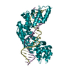
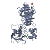
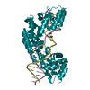
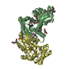
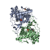
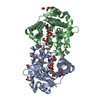
| Title | Crystal Structure of the MTERF1 Y288A substitution bound to the termination sequence. | |||||||||||||||
 Components Components |
| |||||||||||||||
 Keywords Keywords | TRANSCRIPTION/DNA / Protein-DNA / Transcription Factor / Mitochondria / Termination / TRANSCRIPTION-DNA complex | |||||||||||||||
| Function / homology |  Function and homology information Function and homology informationtermination of mitochondrial transcription / Mitochondrial transcription termination / DNA geometric change / mitochondrial nucleoid / Transcriptional activation of mitochondrial biogenesis / DNA-templated transcription termination / double-stranded DNA binding / nucleic acid binding / mitochondrial matrix / mitochondrion ...termination of mitochondrial transcription / Mitochondrial transcription termination / DNA geometric change / mitochondrial nucleoid / Transcriptional activation of mitochondrial biogenesis / DNA-templated transcription termination / double-stranded DNA binding / nucleic acid binding / mitochondrial matrix / mitochondrion / DNA binding / RNA binding Similarity search - Function | |||||||||||||||
| Biological species |  Homo sapiens (human) Homo sapiens (human) | |||||||||||||||
| Method |  X-RAY DIFFRACTION / SYNCHROTRON / MOLECULAR REPLACEMENT / Resolution: 2.65 Å X-RAY DIFFRACTION / SYNCHROTRON / MOLECULAR REPLACEMENT / Resolution: 2.65 Å | |||||||||||||||
| Model details | R162A | |||||||||||||||
 Authors Authors | Byrnes, J. / Hauser, K. / Norona, L. / Mejia, E. / Simmerling, C. / Garcia-Diaz, M. | |||||||||||||||
| Funding support |  United States, 4items United States, 4items
| |||||||||||||||
 Citation Citation | Journal: J.Mol.Biol. / Year: 2016 Title: Base Flipping by MTERF1 Can Accommodate Multiple Conformations and Occurs in a Stepwise Fashion. Authors: Byrnes, J. / Hauser, K. / Norona, L. / Mejia, E. / Simmerling, C. / Garcia-Diaz, M. #1: Journal: Cell / Year: 2010Title: Helix unwinding and base flipping enable human MTERF1 to terminate mitochondrial transcription. Authors: Yakubovskaya, E. / Mejia, E. / Byrnes, J. / Hambardjieva, E. / Garcia-Diaz, M. | |||||||||||||||
| History |
|
- Structure visualization
| Structure viewer | Molecule: MolmilJmol/JSmol |
|---|
- Downloads & links
-Download
| PDBx/mmCIF format | 5co0.cif.gz | 106 KB | Display | PDBx/mmCIF format |
|---|---|---|---|---|
| PDB format | pdb5co0.ent.gz | 75.8 KB | Display | PDB format |
| PDBx/mmJSON format | 5co0.json.gz | Tree view | PDBx/mmJSON format | |
| Others |  Other downloads Other downloads |
-Validation report
| Arichive directory | https://data.pdbj.org/pub/pdb/validation_reports/co/5co0ftp://data.pdbj.org/pub/pdb/validation_reports/co/5co0 | HTTPS FTP |
|---|
-Related structure data
| Related structure data |  5ckyC  5crjC 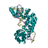 5crkC  3mvaS C: citing same article ( S: Starting model for refinement |
|---|---|
| Similar structure data |


-Links
 PDBj
PDBj


- Assembly
| Deposited unit | 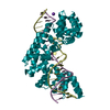
| ||||||||
|---|---|---|---|---|---|---|---|---|---|
| 1 |
| ||||||||
| Unit cell |
|
-Components
| #1: Protein | Mass: 37119.086 Da / Num. of mol.: 1 / Mutation: Y288A Source method: isolated from a genetically manipulated source Source: (gene. exp.) Homo sapiens (human) / Gene: MTERF1 / Plasmid: PTEV-HMBP3 / Production host:  |
|---|---|
| #2: DNA chain | Mass: 6678.315 Da / Num. of mol.: 1 / Source method: obtained synthetically / Source: (synth.) Homo sapiens (human) |
| #3: DNA chain | Mass: 6825.429 Da / Num. of mol.: 1 / Source method: obtained synthetically / Source: (synth.) Homo sapiens (human) |
| #4: Chemical | ChemComp-K /   Mass: 39.098 Da / Num. of mol.: 1 / Source method: obtained synthetically / Formula: K Mass: 39.098 Da / Num. of mol.: 1 / Source method: obtained synthetically / Formula: K |
| #5: Water | ChemComp-HOH /  Mass: 18.015 Da / Num. of mol.: 24 / Source method: isolated from a natural source / Formula: H2O Mass: 18.015 Da / Num. of mol.: 24 / Source method: isolated from a natural source / Formula: H2O |
-Experimental details
-Experiment
| Experiment | Method: X-RAY DIFFRACTION / Number of used crystals: 1 |
|---|
- Sample preparation
| Crystal | Density Matthews: 3.14 Å3/Da / Density % sol: 60.8 % |
|---|---|
| Crystal grow | Temperature: 293 K / Method: vapor diffusion, hanging drop Details: 0.2M Sodium Acetate, 01.M Tris HCl pH 8.0, 15.5% PEG4000 |
-Data collection
| Diffraction | Mean temperature: 100 K | |||||||||||||||||||||||||||
|---|---|---|---|---|---|---|---|---|---|---|---|---|---|---|---|---|---|---|---|---|---|---|---|---|---|---|---|---|
| Diffraction source | Source: SYNCHROTRON / Site: NSLS / Beamline: X9A / Wavelength: 1.075 Å | |||||||||||||||||||||||||||
| Detector | Type: ADSC QUANTUM 315r / Detector: CCD / Date: Oct 11, 2009 | |||||||||||||||||||||||||||
| Radiation | Protocol: SINGLE WAVELENGTH / Monochromatic (M) / Laue (L): M / Scattering type: x-ray | |||||||||||||||||||||||||||
| Radiation wavelength | Wavelength: 1.075 Å / Relative weight: 1 | |||||||||||||||||||||||||||
| Reflection | Resolution: 2.54→80.26 Å / Num. obs: 21454 / % possible obs: 99.9 % / Redundancy: 7.3 % / CC1/2: 1 / Rmerge(I) obs: 0.058 / Rpim(I) all: 0.023 / Net I/σ(I): 26.3 / Num. measured all: 155685 | |||||||||||||||||||||||||||
| Reflection shell | Diffraction-ID: 1 / Rejects: _
|
- Processing
| Software |
| ||||||||||||||||||||||||||||||||||||||||||||||||||||||||||||
|---|---|---|---|---|---|---|---|---|---|---|---|---|---|---|---|---|---|---|---|---|---|---|---|---|---|---|---|---|---|---|---|---|---|---|---|---|---|---|---|---|---|---|---|---|---|---|---|---|---|---|---|---|---|---|---|---|---|---|---|---|---|
| Refinement | Method to determine structure: MOLECULAR REPLACEMENT Starting model: 3MVA Resolution: 2.65→80.26 Å / Cor.coef. Fo:Fc: 0.943 / Cor.coef. Fo:Fc free: 0.918 / SU B: 11.368 / SU ML: 0.235 / Cross valid method: THROUGHOUT / σ(F): 0 / ESU R: 0.53 / ESU R Free: 0.315 / Stereochemistry target values: MAXIMUM LIKELIHOOD Details: HYDROGENS HAVE BEEN ADDED IN THE RIDING POSITIONS U VALUES : REFINED INDIVIDUALLY
| ||||||||||||||||||||||||||||||||||||||||||||||||||||||||||||
| Solvent computation | Ion probe radii: 0.8 Å / Shrinkage radii: 0.8 Å / VDW probe radii: 1.2 Å / Solvent model: MASK | ||||||||||||||||||||||||||||||||||||||||||||||||||||||||||||
| Displacement parameters | Biso max: 136.36 Å2 / Biso mean: 59.248 Å2 / Biso min: 25.38 Å2
| ||||||||||||||||||||||||||||||||||||||||||||||||||||||||||||
| Refinement step | Cycle: final / Resolution: 2.65→80.26 Å
| ||||||||||||||||||||||||||||||||||||||||||||||||||||||||||||
| Refine LS restraints |
| ||||||||||||||||||||||||||||||||||||||||||||||||||||||||||||
| LS refinement shell | Resolution: 2.65→2.717 Å / Total num. of bins used: 20
|
