[English] 日本語
Yorodumi
- PDB-5cek: Pseudokinase domain of Human Tribbles Homolog 1 -

+
Open data


ID or keywords:

Loading...

-
Basic information

Entry
Database: PDB / ID: 5cek
TitlePseudokinase domain of Human Tribbles Homolog 1
ComponentsTribbles homolog 1
KeywordsTRANSFERASE / kinase
Function / homology
Function and homology information


ubiquitin-protein transferase regulator activity / positive regulation of eosinophil differentiation / negative regulation of smooth muscle cell migration / regulation of MAP kinase activity / negative regulation of neutrophil differentiation / positive regulation of macrophage differentiation / negative regulation of lipopolysaccharide-mediated signaling pathway / NGF-stimulated transcription / mitogen-activated protein kinase kinase binding / protein kinase inhibitor activity ...ubiquitin-protein transferase regulator activity / positive regulation of eosinophil differentiation / negative regulation of smooth muscle cell migration / regulation of MAP kinase activity / negative regulation of neutrophil differentiation / positive regulation of macrophage differentiation / negative regulation of lipopolysaccharide-mediated signaling pathway / NGF-stimulated transcription / mitogen-activated protein kinase kinase binding / protein kinase inhibitor activity / JNK cascade / negative regulation of smooth muscle cell proliferation / negative regulation of protein kinase activity / negative regulation of DNA-binding transcription factor activity / positive regulation of proteasomal ubiquitin-dependent protein catabolic process / RNA polymerase II-specific DNA-binding transcription factor binding / response to lipopolysaccharide / ubiquitin protein ligase binding / nucleus / cytosol / cytoplasm
Similarity search - Function
Tribbles homologue 1 / Pseudokinase tribbles family/serine-threonine-protein kinase 40 / Transferase(Phosphotransferase) domain 1 / Transferase(Phosphotransferase); domain 1 / Phosphorylase Kinase; domain 1 / Phosphorylase Kinase; domain 1 / Protein kinase domain / Serine/Threonine protein kinases, catalytic domain / Protein kinase domain profile. / Protein kinase domain ...Tribbles homologue 1 / Pseudokinase tribbles family/serine-threonine-protein kinase 40 / Transferase(Phosphotransferase) domain 1 / Transferase(Phosphotransferase); domain 1 / Phosphorylase Kinase; domain 1 / Phosphorylase Kinase; domain 1 / Protein kinase domain / Serine/Threonine protein kinases, catalytic domain / Protein kinase domain profile. / Protein kinase domain / Protein kinase-like domain superfamily / 2-Layer Sandwich / Orthogonal Bundle / Mainly Alpha / Alpha Beta
Similarity search - Domain/homology
Biological speciesHomo sapiens (human)
MethodX-RAY DIFFRACTION / SYNCHROTRON / Resolution: 2.8 Å
AuthorsMace, P.D. / Nakatani, Y.
Funding support New Zealand, 2items
OrganizationGrant numberCountry
Royal Society of New Zealand New Zealand
Health Research Council (HRC) New Zealand
CitationJournal: Structure / Year: 2015
Title: Molecular Mechanism of CCAAT-Enhancer Binding Protein Recruitment by the TRIB1 Pseudokinase.
Authors: Murphy, J.M. / Nakatani, Y. / Jamieson, S.A. / Dai, W. / Lucet, I.S. / Mace, P.D.
History
DepositionJul 7, 2015Deposition site: RCSB / Processing site: RCSB
Revision 1.0Nov 11, 2015Provider: repository / Type: Initial release
Revision 1.1Nov 18, 2015Group: Database references
Revision 1.2Sep 20, 2017Group: Data collection / Database references / Derived calculations
Category: citation / diffrn_source / pdbx_struct_oper_list
Item: _citation.journal_id_CSD / _diffrn_source.pdbx_synchrotron_site / _pdbx_struct_oper_list.symmetry_operation
Revision 1.3Jan 1, 2020Group: Author supporting evidence / Category: pdbx_audit_support / Item: _pdbx_audit_support.funding_organization
Revision 1.4Mar 6, 2024Group: Data collection / Database references / Category: chem_comp_atom / chem_comp_bond / database_2
Item: _database_2.pdbx_DOI / _database_2.pdbx_database_accession

-
Structure visualization

Structure viewerMolecule:
MolmilJmol/JSmol

Downloads & links

-
Assembly

Deposited unit
A: Tribbles homolog 1


Theoretical massNumber of molelcules
Total (without water)29,9561
Polymers29,9561
Non-polymers00
Water0
1


  • Idetical with deposited unit
  • defined by author&software
TypeNameSymmetry operationNumber
identity operation1_555x,y,z1
MethodPISA
Unit cell
Length a, b, c (Å)152.693, 152.693, 62.798
Angle α, β, γ (deg.)90.00, 90.00, 120.00
Int Tables number180
Space group name H-MP6222

-
Components

#1: Protein Tribbles homolog 1 / TRB-1 / G-protein-coupled receptor-induced gene 2 protein / GIG-2 / SKIP1


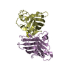
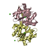
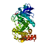
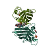
Mass: 29955.529 Da / Num. of mol.: 1 / Fragment: unp residues 83-343
Source method: isolated from a genetically manipulated source
Source: (gene. exp.) Homo sapiens (human) / Gene: TRIB1, C8FW, GIG2, TRB1 / Production host: Escherichia coli (E. coli) / Strain (production host): BL21(DE3) / References: UniProt: Q96RU8

-
Experimental details

-
Experiment

ExperimentMethod: X-RAY DIFFRACTION

-
Sample preparation

CrystalDensity Matthews: 3.53 Å3/Da / Density % sol: 65.13 %
Crystal growTemperature: 290 K / Method: vapor diffusion, sitting drop / pH: 5.5
Details: 0.1 M Bis-Tris, 2-3 M NaCl, 0.1 M Sodium Citrate and 0.01 M reduced L-Glutathione, 0.01 M oxidized L-Glutathione

-
Data collection

DiffractionMean temperature: 100 K
Diffraction sourceSource: SYNCHROTRON / Site: Australian Synchrotron / Beamline: MX1 / Wavelength: 0.954 Å
DetectorType: ADSC QUANTUM 210r / Detector: CCD / Date: May 2, 2014
RadiationProtocol: SINGLE WAVELENGTH / Monochromatic (M) / Laue (L): M / Scattering type: x-ray
Radiation wavelengthWavelength: 0.954 Å / Relative weight: 1
ReflectionResolution: 2.8→48.5 Å / Num. obs: 11093 / % possible obs: 100 % / Redundancy: 9.6 % / Rmerge(I) obs: 0.097 / Net I/σ(I): 18.6
Reflection shellResolution: 2.8→2.95 Å / Redundancy: 9.9 % / Mean I/σ(I) obs: 2 / % possible all: 100

-
Processing

Software
NameVersionClassification
REFMAC5.8.0107refinement
XDSdata reduction
SCALAdata scaling
SHELXDEphasing
RefinementResolution: 2.8→48.5 Å / Cor.coef. Fo:Fc: 0.957 / Cor.coef. Fo:Fc free: 0.938 / SU B: 39.215 / SU ML: 0.299 / Cross valid method: THROUGHOUT / ESU R: 0.489 / ESU R Free: 0.3 / Stereochemistry target values: MAXIMUM LIKELIHOOD / Details: HYDROGENS HAVE BEEN ADDED IN THE RIDING POSITIONS
RfactorNum. reflection% reflectionSelection details
Rfree0.24554 590 5.3 %RANDOM
Rwork0.20736 ---
obs0.20937 10489 99.89 %-
Solvent computationIon probe radii: 0.8 Å / Shrinkage radii: 0.8 Å / VDW probe radii: 1.1 Å / Solvent model: MASK
Displacement parametersBiso mean: 100.759 Å2
Baniso -1Baniso -2Baniso -3
1-2.12 Å21.06 Å2-0 Å2
2--2.12 Å2-0 Å2
3----6.88 Å2
Refinement stepCycle: 1 / Resolution: 2.8→48.5 Å
ProteinNucleic acidLigandSolventTotal
Num. atoms1919 0 0 0 1919
Refine LS restraints
Refine-IDTypeDev idealDev ideal targetNumber
X-RAY DIFFRACTIONr_bond_refined_d0.0080.0191962
X-RAY DIFFRACTIONr_bond_other_d0.0020.021926
X-RAY DIFFRACTIONr_angle_refined_deg1.3761.9712647
X-RAY DIFFRACTIONr_angle_other_deg0.98234423
X-RAY DIFFRACTIONr_dihedral_angle_1_deg7.3565232
X-RAY DIFFRACTIONr_dihedral_angle_2_deg33.47721.49487
X-RAY DIFFRACTIONr_dihedral_angle_3_deg14.76915349
X-RAY DIFFRACTIONr_dihedral_angle_4_deg14.6541521
X-RAY DIFFRACTIONr_chiral_restr0.0710.2298
X-RAY DIFFRACTIONr_gen_planes_refined0.0050.0212119
X-RAY DIFFRACTIONr_gen_planes_other0.0020.02462
X-RAY DIFFRACTIONr_nbd_refined
X-RAY DIFFRACTIONr_nbd_other
X-RAY DIFFRACTIONr_nbtor_refined
X-RAY DIFFRACTIONr_nbtor_other
X-RAY DIFFRACTIONr_xyhbond_nbd_refined
X-RAY DIFFRACTIONr_xyhbond_nbd_other
X-RAY DIFFRACTIONr_metal_ion_refined
X-RAY DIFFRACTIONr_metal_ion_other
X-RAY DIFFRACTIONr_symmetry_vdw_refined
X-RAY DIFFRACTIONr_symmetry_vdw_other
X-RAY DIFFRACTIONr_symmetry_hbond_refined
X-RAY DIFFRACTIONr_symmetry_hbond_other
X-RAY DIFFRACTIONr_symmetry_metal_ion_refined
X-RAY DIFFRACTIONr_symmetry_metal_ion_other
X-RAY DIFFRACTIONr_mcbond_it5.5255.64943
X-RAY DIFFRACTIONr_mcbond_other5.5225.639942
X-RAY DIFFRACTIONr_mcangle_it7.9218.441170
X-RAY DIFFRACTIONr_mcangle_other7.9188.4411171
X-RAY DIFFRACTIONr_scbond_it6.356.3371019
X-RAY DIFFRACTIONr_scbond_other6.3466.3371020
X-RAY DIFFRACTIONr_scangle_it
X-RAY DIFFRACTIONr_scangle_other9.4819.2581478
X-RAY DIFFRACTIONr_long_range_B_refined13.47754.5637948
X-RAY DIFFRACTIONr_long_range_B_other13.47654.5647949
X-RAY DIFFRACTIONr_rigid_bond_restr
X-RAY DIFFRACTIONr_sphericity_free
X-RAY DIFFRACTIONr_sphericity_bonded
LS refinement shellResolution: 2.8→2.873 Å / Total num. of bins used: 20
RfactorNum. reflection% reflection
Rfree0.403 47 -
Rwork0.327 750 -
obs--100 %
Refinement TLS params.Method: refined / Origin x: -33.661 Å / Origin y: 36.677 Å / Origin z: -0.76 Å
111213212223313233
T0.3624 Å2-0.1539 Å2-0.0023 Å2-0.0841 Å2-0.0419 Å2--0.2055 Å2
L2.4209 °2-0.2853 °20.2204 °2-5.7178 °2-2.3697 °2--5.4594 °2
S0.1563 Å °-0.1638 Å °0.5347 Å °0.8336 Å °-0.1235 Å °-0.522 Å °0.1863 Å °-0.2869 Å °-0.0329 Å °

+
About Yorodumi

-
News

-
Feb 9, 2022. New format data for meta-information of EMDB entries

New format data for meta-information of EMDB entries

  • Version 3 of the EMDB header file is now the official format.
  • The previous official version 1.9 will be removed from the archive.

Related info.:EMDB header

External links:wwPDB to switch to version 3 of the EMDB data model

-
Aug 12, 2020. Covid-19 info

Covid-19 info

URL: https://pdbj.org/emnavi/covid19.php

New page: Covid-19 featured information page in EM Navigator.

Related info.:Covid-19 info / Mar 5, 2020. Novel coronavirus structure data

+
Mar 5, 2020. Novel coronavirus structure data

Novel coronavirus structure data

Related info.:Yorodumi Speices / Aug 12, 2020. Covid-19 info

External links:COVID-19 featured content - PDBj / Molecule of the Month (242):Coronavirus Proteases

+
Jan 31, 2019. EMDB accession codes are about to change! (news from PDBe EMDB page)

EMDB accession codes are about to change! (news from PDBe EMDB page)

  • The allocation of 4 digits for EMDB accession codes will soon come to an end. Whilst these codes will remain in use, new EMDB accession codes will include an additional digit and will expand incrementally as the available range of codes is exhausted. The current 4-digit format prefixed with “EMD-” (i.e. EMD-XXXX) will advance to a 5-digit format (i.e. EMD-XXXXX), and so on. It is currently estimated that the 4-digit codes will be depleted around Spring 2019, at which point the 5-digit format will come into force.
  • The EM Navigator/Yorodumi systems omit the EMD- prefix.

Related info.:Q: What is EMD? / ID/Accession-code notation in Yorodumi/EM Navigator

External links:EMDB Accession Codes are Changing Soon! / Contact to PDBj

+
Jul 12, 2017. Major update of PDB

Major update of PDB

  • wwPDB released updated PDB data conforming to the new PDBx/mmCIF dictionary.
  • This is a major update changing the version number from 4 to 5, and with Remediation, in which all the entries are updated.
  • In this update, many items about electron microscopy experimental information are reorganized (e.g. em_software).
  • Now, EM Navigator and Yorodumi are based on the updated data.

External links:wwPDB Remediation / Enriched Model Files Conforming to OneDep Data Standards Now Available in the PDB FTP Archive

-
Yorodumi

Thousand views of thousand structures

  • Yorodumi is a browser for structure data from EMDB, PDB, SASBDB, etc.
  • This page is also the successor to EM Navigator detail page, and also detail information page/front-end page for Omokage search.
  • The word "yorodu" (or yorozu) is an old Japanese word meaning "ten thousand". "mi" (miru) is to see.

Related info.:EMDB / PDB / SASBDB / Comparison of 3 databanks / Yorodumi Search / Aug 31, 2016. New EM Navigator & Yorodumi / Yorodumi Papers / Jmol/JSmol / Function and homology information / Changes in new EM Navigator and Yorodumi

Read more