Mass: 18.015 Da / Num. of mol.: 46 / Source method: isolated from a natural source / Formula: H2O
Has protein modification
Y
-
Experimental details
-
Experiment
Experiment
Method: X-RAY DIFFRACTION
-
Sample preparation
Crystal
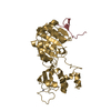
Density Matthews: 2.06 Å3/Da / Density % sol: 40.19 % / Description: Crystals were grown as long rods
Crystal grow
Temperature: 293 K / Method: vapor diffusion, sitting drop / pH: 6 Details: Activated hepsin was incubated with 10 mM Benzamidine at 4 oC for 2 hours and concentrated to7 mg/ml using 10 KDa cut off centricon [Millipore]. 1microlit of protein was mixed with 1 ...Details: Activated hepsin was incubated with 10 mM Benzamidine at 4 oC for 2 hours and concentrated to7 mg/ml using 10 KDa cut off centricon [Millipore]. 1microlit of protein was mixed with 1 microlit of reservoir buffer containing 100 mM sodium Cacodylate (6.0), 22% PEG 3350, and 200 mM ammonium fluoride. Plate clusters with a dimension of 50 micron m x 100 micron m were observed after 2 days of crystallization set up. PH range: 5.8 - 6.3
Resolution: 2.5→58.63 Å / Cor.coef. Fo:Fc: 0.935 / Cor.coef. Fo:Fc free: 0.859 / SU B: 10.623 / SU ML: 0.246 / Cross valid method: THROUGHOUT / ESU R Free: 0.399 / Stereochemistry target values: MAXIMUM LIKELIHOOD Details: THE REASON FOR POOR ELECTRON DENSITY IS THAT THE REGION BETWEEN A LEU 158 AND RESIDUE A ILE 163 HIGHLY DISORDERED AND HENCE THE ATOMS POSITIONS ARE NOT STABILIZED IN THE CRYSTAL. HYDROGENS ...Details: THE REASON FOR POOR ELECTRON DENSITY IS THAT THE REGION BETWEEN A LEU 158 AND RESIDUE A ILE 163 HIGHLY DISORDERED AND HENCE THE ATOMS POSITIONS ARE NOT STABILIZED IN THE CRYSTAL. HYDROGENS HAVE BEEN ADDED IN THE RIDING POSITIONS
Rfactor
Num. reflection
% reflection
Selection details
Rfree
0.2815
485
4.8 %
RANDOM
Rwork
0.18714
-
-
-
obs
0.19165
9554
86.66 %
-
Solvent computation
Ion probe radii: 0.8 Å / Shrinkage radii: 0.8 Å / VDW probe radii: 1.2 Å / Solvent model: MASK
 Movie
Movie Controller

 Yorodumi
 Open data
 Basic information
 Components
 Keywords
 Function and homology information
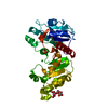
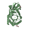
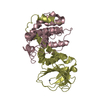
 Homo sapiens (human)
 X-RAY DIFFRACTION /
  Authors
 Citation
 Structure visualization
 Downloads & links
 Other downloads
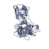
 PDBj
PDBj Assembly
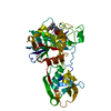

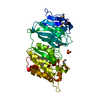
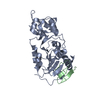
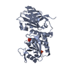
 SPODOPTERA FRUGIPERDA (fall armyworm) / References: UniProt: P05981, hepsin
 Mass: 334.415 Da / Num. of mol.: 1 / Source method: obtained synthetically / Formula: C20H22N4O
 Mass: 18.015 Da / Num. of mol.: 46 / Source method: isolated from a natural source / Formula: H2O
 Sample preparation
 Processing