 ムービー
ムービー コントローラー


+ データを開く

- 基本情報
基本情報
| 登録情報 | データベース: PDB / ID: 5bzh | ||||||
|---|---|---|---|---|---|---|---|
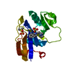
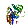
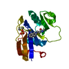
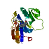
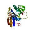
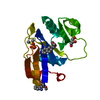
| タイトル | Crystal structure of the murine CD44 hyaluronan binding domain complex with a small molecule | ||||||
 要素 要素 | CD44 antigen | ||||||
 キーワード キーワード | PROTEIN BINDING / Link module | ||||||
| 機能・相同性 |  機能・相同性情報 機能・相同性情報Hyaluronan uptake and degradation / macrophage fusion / hyaluronic acid binding / macrophage migration inhibitory factor receptor complex / negative regulation of regulatory T cell differentiation / Degradation of the extracellular matrix / regulation of lamellipodium morphogenesis / Integrin cell surface interactions / Cell surface interactions at the vascular wall / hyaluronan catabolic process ...Hyaluronan uptake and degradation / macrophage fusion / hyaluronic acid binding / macrophage migration inhibitory factor receptor complex / negative regulation of regulatory T cell differentiation / Degradation of the extracellular matrix / regulation of lamellipodium morphogenesis / Integrin cell surface interactions / Cell surface interactions at the vascular wall / hyaluronan catabolic process / wound healing involved in inflammatory response / positive regulation of adaptive immune response / positive regulation of neutrophil apoptotic process / branching involved in prostate gland morphogenesis / type II transforming growth factor beta receptor binding / negative regulation of mature B cell apoptotic process / negative regulation of CD4-positive, alpha-beta T cell proliferation / cargo receptor activity / wound healing, spreading of cells / epidermal growth factor receptor binding / branching involved in ureteric bud morphogenesis / negative regulation of intrinsic apoptotic signaling pathway in response to DNA damage by p53 class mediator / channel regulator activity / negative regulation of DNA damage response, signal transduction by p53 class mediator / lamellipodium membrane / microvillus / Neutrophil degranulation / receptor-mediated endocytosis / cell projection / regulation of cell growth / phosphoprotein binding / Wnt signaling pathway / cytokine-mediated signaling pathway / negative regulation of inflammatory response / positive regulation of peptidyl-tyrosine phosphorylation / transmembrane signaling receptor activity / neuron projection development / cell migration / positive regulation of peptidyl-serine phosphorylation / basolateral plasma membrane / positive regulation of ERK1 and ERK2 cascade / cell adhesion / inflammatory response / apical plasma membrane / membrane raft / external side of plasma membrane / positive regulation of gene expression / protein kinase binding / cell surface / protein-containing complex / extracellular region / plasma membrane 類似検索 - 分子機能 | ||||||
| 生物種 |  | ||||||
| 手法 |  X線回折 / 分子置換 / 解像度: 1.95 Å X線回折 / 分子置換 / 解像度: 1.95 Å | ||||||
 データ登録者 データ登録者 | Liu, L.K. / Finzel, B.C. | ||||||
 引用 引用 | ジャーナル: To Be Published タイトル: Crystal structure of the murine cd44 hyaluronan binding domain complex with a small molecule 著者: Liu, L.K. / Finzel, B.C. | ||||||
| 履歴 |
|
- 構造の表示
構造の表示
| 構造ビューア | 分子: MolmilJmol/JSmol |
|---|
- ダウンロードとリンク
-ダウンロード
| PDBx/mmCIF形式 | 5bzh.cif.gz | 41.6 KB | 表示 | PDBx/mmCIF形式 |
|---|---|---|---|---|
| PDB形式 | pdb5bzh.ent.gz | 29.5 KB | 表示 | PDB形式 |
| PDBx/mmJSON形式 | 5bzh.json.gz | ツリー表示 | PDBx/mmJSON形式 | |
| その他 |  その他のダウンロード その他のダウンロード |
-検証レポート
| 文書・要旨 | 5bzh_validation.pdf.gz | 443.9 KB | 表示 | wwPDB検証レポート |
|---|---|---|---|---|
| 文書・詳細版 | 5bzh_full_validation.pdf.gz | 443.9 KB | 表示 | |
| XML形式データ | 5bzh_validation.xml.gz | 8.4 KB | 表示 | |
| CIF形式データ | 5bzh_validation.cif.gz | 10.7 KB | 表示 | |
| アーカイブディレクトリ | https://data.pdbj.org/pub/pdb/validation_reports/bz/5bzhftp://data.pdbj.org/pub/pdb/validation_reports/bz/5bzh | HTTPS FTP |
-関連構造データ
| 関連構造データ |  5bzcC 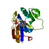 5bzeC  5bzfC 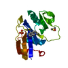 5bzgC 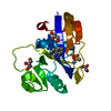 5bziC  5bzjC  5bzkC  5bzlC 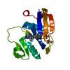 5bzmC 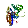 5bznC  5bzoC  5bzpC 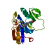 5bzqC  5bzrC 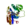 5bzsC  5bztC C: 同じ文献を引用 ( |
|---|---|
| 類似構造データ |


-リンク
 PDBj
PDBj


- 集合体
集合体
| 登録構造単位 | 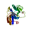
| ||||||||
|---|---|---|---|---|---|---|---|---|---|
| 1 |
| ||||||||
| 単位格子 |
|
-要素
| #1: タンパク質 | 分子量: 16855.803 Da / 分子数: 1 / 変異: H23M, Q24N / 由来タイプ: 組換発現 / 由来: (組換発現)  |
|---|---|
| #2: 化合物 | ChemComp-DMS /   分子量: 78.133 Da / 分子数: 1 / 由来タイプ: 合成 / 式: C2H6OS / コメント: DMSO, 沈殿剤*YM 分子量: 78.133 Da / 分子数: 1 / 由来タイプ: 合成 / 式: C2H6OS / コメント: DMSO, 沈殿剤*YM |
| #3: 化合物 | ChemComp-4X1 /   分子量: 190.285 Da / 分子数: 1 / 由来タイプ: 合成 / 式: C12H18N2 分子量: 190.285 Da / 分子数: 1 / 由来タイプ: 合成 / 式: C12H18N2 |
| #4: 水 | ChemComp-HOH /  分子量: 18.015 Da / 分子数: 53 / 由来タイプ: 天然 / 式: H2O 分子量: 18.015 Da / 分子数: 53 / 由来タイプ: 天然 / 式: H2O |
-実験情報
-実験
| 実験 | 手法: X線回折 / 使用した結晶の数: 1 |
|---|
- 試料調製
試料調製
| 結晶 | マシュー密度: 2.14 Å3/Da / 溶媒含有率: 42.47 % |
|---|---|
| 結晶化 | 温度: 298 K / 手法: 蒸気拡散法, ハンギングドロップ法 / pH: 6.5 / 詳細: 30% PEG MME 5000, 100 mM MES, 200 mM (NH4)2SO4 |
-データ収集
| 回折 | 平均測定温度: 100 K | ||||||||||||||||||||||||||||||||||||||||||||||||||||||||||||||||||||||||||||||||||||||||||||||||||||||||||||||||||||||||||||||||||||||||||||||||||||||||||||||||||||||||||||||||||||||||||||||||||||||||||||||||||
|---|---|---|---|---|---|---|---|---|---|---|---|---|---|---|---|---|---|---|---|---|---|---|---|---|---|---|---|---|---|---|---|---|---|---|---|---|---|---|---|---|---|---|---|---|---|---|---|---|---|---|---|---|---|---|---|---|---|---|---|---|---|---|---|---|---|---|---|---|---|---|---|---|---|---|---|---|---|---|---|---|---|---|---|---|---|---|---|---|---|---|---|---|---|---|---|---|---|---|---|---|---|---|---|---|---|---|---|---|---|---|---|---|---|---|---|---|---|---|---|---|---|---|---|---|---|---|---|---|---|---|---|---|---|---|---|---|---|---|---|---|---|---|---|---|---|---|---|---|---|---|---|---|---|---|---|---|---|---|---|---|---|---|---|---|---|---|---|---|---|---|---|---|---|---|---|---|---|---|---|---|---|---|---|---|---|---|---|---|---|---|---|---|---|---|---|---|---|---|---|---|---|---|---|---|---|---|---|---|---|---|---|
| 放射光源 | 由来: 回転陽極 / タイプ: RIGAKU MICROMAX-007 HF / 波長: 1.5418 Å | ||||||||||||||||||||||||||||||||||||||||||||||||||||||||||||||||||||||||||||||||||||||||||||||||||||||||||||||||||||||||||||||||||||||||||||||||||||||||||||||||||||||||||||||||||||||||||||||||||||||||||||||||||
| 検出器 | タイプ: RIGAKU SATURN 944+ / 検出器: CCD / 日付: 2013年11月12日 | ||||||||||||||||||||||||||||||||||||||||||||||||||||||||||||||||||||||||||||||||||||||||||||||||||||||||||||||||||||||||||||||||||||||||||||||||||||||||||||||||||||||||||||||||||||||||||||||||||||||||||||||||||
| 放射 | プロトコル: SINGLE WAVELENGTH / 単色(M)・ラウエ(L): M / 散乱光タイプ: x-ray | ||||||||||||||||||||||||||||||||||||||||||||||||||||||||||||||||||||||||||||||||||||||||||||||||||||||||||||||||||||||||||||||||||||||||||||||||||||||||||||||||||||||||||||||||||||||||||||||||||||||||||||||||||
| 放射波長 | 波長: 1.5418 Å / 相対比: 1 | ||||||||||||||||||||||||||||||||||||||||||||||||||||||||||||||||||||||||||||||||||||||||||||||||||||||||||||||||||||||||||||||||||||||||||||||||||||||||||||||||||||||||||||||||||||||||||||||||||||||||||||||||||
| 反射 | 解像度: 1.95→8.72 Å / Num. obs: 9978 / % possible obs: 96.3 % / Observed criterion σ(I): -3 / 冗長度: 3.753 % / Biso Wilson estimate: 15.378 Å2 / Rmerge F obs: 1 / Rmerge(I) obs: 0.017 / Rrim(I) all: 0.02 / Χ2: 0.99 / Net I/σ(I): 56.83 / Num. measured all: 37452 | ||||||||||||||||||||||||||||||||||||||||||||||||||||||||||||||||||||||||||||||||||||||||||||||||||||||||||||||||||||||||||||||||||||||||||||||||||||||||||||||||||||||||||||||||||||||||||||||||||||||||||||||||||
| 反射 シェル | Diffraction-ID: 1 / Rejects: _
|
-位相決定
| 位相決定 | 手法: 分子置換 |
|---|
- 解析
解析
| ソフトウェア |
| |||||||||||||||||||||||||||||||||||||||||||||||||||||||||||||||||
|---|---|---|---|---|---|---|---|---|---|---|---|---|---|---|---|---|---|---|---|---|---|---|---|---|---|---|---|---|---|---|---|---|---|---|---|---|---|---|---|---|---|---|---|---|---|---|---|---|---|---|---|---|---|---|---|---|---|---|---|---|---|---|---|---|---|---|
| 精密化 | 構造決定の手法: 分子置換 / 解像度: 1.95→8.72 Å / Cor.coef. Fo:Fc: 0.938 / Cor.coef. Fo:Fc free: 0.882 / WRfactor Rfree: 0.2181 / WRfactor Rwork: 0.167 / FOM work R set: 0.8629 / SU B: 3.338 / SU ML: 0.099 / SU R Cruickshank DPI: 0.183 / SU Rfree: 0.1666 / 交差検証法: THROUGHOUT / σ(F): 0 / ESU R: 0.183 / ESU R Free: 0.167 / 立体化学のターゲット値: MAXIMUM LIKELIHOOD 詳細: HYDROGENS HAVE BEEN ADDED IN THE RIDING POSITIONS U VALUES : REFINED INDIVIDUALLY
| |||||||||||||||||||||||||||||||||||||||||||||||||||||||||||||||||
| 溶媒の処理 | イオンプローブ半径: 0.8 Å / 減衰半径: 0.8 Å / VDWプローブ半径: 1.4 Å / 溶媒モデル: MASK | |||||||||||||||||||||||||||||||||||||||||||||||||||||||||||||||||
| 原子変位パラメータ | Biso max: 37.44 Å2 / Biso mean: 9.05 Å2 / Biso min: 2 Å2
| |||||||||||||||||||||||||||||||||||||||||||||||||||||||||||||||||
| 精密化ステップ | サイクル: final / 解像度: 1.95→8.72 Å
| |||||||||||||||||||||||||||||||||||||||||||||||||||||||||||||||||
| 拘束条件 |
| |||||||||||||||||||||||||||||||||||||||||||||||||||||||||||||||||
| LS精密化 シェル | 解像度: 1.951→2.001 Å / Total num. of bins used: 20
|
