Mass: 18.015 Da / Num. of mol.: 243 / Source method: isolated from a natural source / Formula: H2O
Has protein modification
Y
Sequence details
THE CONSTRUCT (26-186) WAS EXPRESSED WITH A PURIFICATION TAG MGSDKIHHHHHHENLYFQG. THE TAG WAS ...THE CONSTRUCT (26-186) WAS EXPRESSED WITH A PURIFICATION TAG MGSDKIHHHHHHENLYFQG. THE TAG WAS REMOVED WITH TEV PROTEASE LEAVING ONLY A GLYCINE (0) FOLLOWED BY THE TARGET SEQUENCE.
-
Experimental details
-
Experiment
Experiment
Method: X-RAY DIFFRACTION / Number of used crystals: 1
-
Sample preparation
Crystal
Density Matthews: 3.26 Å3/Da / Density % sol: 62.25 %
Crystal grow
Temperature: 277 K / Method: vapor diffusion, sitting drop Details: 20% polyethylene glycol 3350, 0.2M tri-sodium citrate
Type: DECTRIS PILATUS 6M / Detector: PIXEL / Date: May 15, 2015 Details: Flat mirror (vertical focusing); single crystal Si(111) bent monochromator (horizontal focusing)
Radiation
Monochromator: single crystal Si(111) bent / Protocol: SINGLE WAVELENGTH / Monochromatic (M) / Laue (L): M / Scattering type: x-ray
Radiation wavelength
Wavelength: 0.97923 Å / Relative weight: 1
Reflection
Resolution: 1.9→49.453 Å / Num. obs: 18773 / % possible obs: 99.7 % / Observed criterion σ(I): -3 / Redundancy: 6.253 % / Biso Wilson estimate: 32.52 Å2 / Rmerge F obs: 0.999 / Rmerge(I) obs: 0.065 / Rrim(I) all: 0.071 / Net I/σ(I): 15.17 / Num. measured all: 117394
Reflection shell
Resolution (Å)
Rmerge F obs
Rmerge(I) obs
Mean I/σ(I) obs
Num. measured obs
Num. possible
Num. unique obs
Rrim(I) all
Diffraction-ID
% possible all
1.9-1.97
0.689
0.615
1.7
6442
1945
1909
0.73
1
98.1
1.97-2.05
0.894
0.492
3.3
11871
1876
1874
0.536
99.9
2.05-2.14
0.945
0.341
4.9
12021
1797
1793
0.37
99.8
2.14-2.25
0.978
0.227
7.2
12659
1830
1830
0.245
100
2.25-2.39
0.985
0.167
9.6
12579
1877
1877
0.181
100
2.39-2.58
0.99
0.132
11.9
12376
1913
1913
0.143
100
2.58-2.84
0.995
0.095
17.3
13042
1884
1884
0.103
100
2.84-3.25
0.997
0.062
24.7
11981
1875
1873
0.068
99.9
3.25-4.08
0.998
0.046
33.2
12297
1887
1883
0.05
99.8
4.08-49.453
0.999
0.04
36.8
12126
1943
1937
0.044
99.7
-
Phasing
Phasing
Method: SAD
-
Processing
Software
Name
Version
Classification
PDB_EXTRACT
3.1
dataextraction
XDS
November3, 2014BUILT=20141118
datascaling
XSCALE
datascaling
SHELX
phasing
SHARP
phasing
SHELXD
phasing
BUSTER
2.10.2
refinement
Refinement
Method to determine structure: SAD / Resolution: 1.9→49.453 Å / Cor.coef. Fo:Fc: 0.9618 / Cor.coef. Fo:Fc free: 0.9525 / Occupancy max: 1 / Occupancy min: 0.3 / Cross valid method: THROUGHOUT / σ(F): 0 Details: 1. A MET-INHIBITION PROTOCOL WAS USED FOR SELENOMETHIONINE INCORPORATION DURING PROTEIN EXPRESSION. THE OCCUPANCY OF THE SE ATOMS IN THE MSE RESIDUES WAS REDUCED TO 0.75 FOR THE REDUCED ...Details: 1. A MET-INHIBITION PROTOCOL WAS USED FOR SELENOMETHIONINE INCORPORATION DURING PROTEIN EXPRESSION. THE OCCUPANCY OF THE SE ATOMS IN THE MSE RESIDUES WAS REDUCED TO 0.75 FOR THE REDUCED SCATTERING POWER DUE TO PARTIAL S-MET INCORPORATION. 2. THE SAD PHASES WERE USED AS RESTRAINTS DURING REFINEMENT. 3. ATOM RECORDS CONTAIN SUM OF TLS AND RESIDUAL B FACTORS. ANISOU RECORDS CONTAIN SUM OF TLS AND RESIDUAL U FACTORS.
In the structure databanks used in Yorodumi, some data are registered as the other names, "COVID-19 virus" and "2019-nCoV". Here are the details of the virus and the list of structure data.
Jan 31, 2019. EMDB accession codes are about to change! (news from PDBe EMDB page)
EMDB accession codes are about to change! (news from PDBe EMDB page)
The allocation of 4 digits for EMDB accession codes will soon come to an end. Whilst these codes will remain in use, new EMDB accession codes will include an additional digit and will expand incrementally as the available range of codes is exhausted. The current 4-digit format prefixed with “EMD-” (i.e. EMD-XXXX) will advance to a 5-digit format (i.e. EMD-XXXXX), and so on. It is currently estimated that the 4-digit codes will be depleted around Spring 2019, at which point the 5-digit format will come into force.
The EM Navigator/Yorodumi systems omit the EMD- prefix.
Related info.:Q: What is EMD? / ID/Accession-code notation in Yorodumi/EM Navigator
Yorodumi is a browser for structure data from EMDB, PDB, SASBDB, etc.
This page is also the successor to EM Navigator detail page, and also detail information page/front-end page for Omokage search.
The word "yorodu" (or yorozu) is an old Japanese word meaning "ten thousand". "mi" (miru) is to see.
Related info.:EMDB / PDB / SASBDB / Comparison of 3 databanks / Yorodumi Search / Aug 31, 2016. New EM Navigator & Yorodumi / Yorodumi Papers / Jmol/JSmol / Function and homology information / Changes in new EM Navigator and Yorodumi
 Movie
Movie Controller

 Yorodumi
 Open data
 Basic information
 Components
 Keywords
 Function and homology information
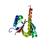
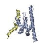
 Peptoclostridium difficile (bacteria)
 X-RAY DIFFRACTION /
  Authors
 Citation
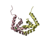
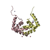
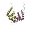
 Structure visualization
 Downloads & links
 Other downloads

 PDBj
PDBj


 Assembly
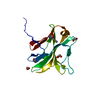

 Mass: 62.068 Da / Num. of mol.: 3 / Source method: obtained synthetically / Formula: C2H6O2
 Mass: 18.015 Da / Num. of mol.: 243 / Source method: isolated from a natural source / Formula: H2O
 Sample preparation
 / Beamline: BL11-1 / Wavelength: 0.97923 Å
 Processing