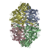
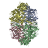
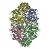
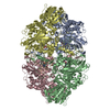
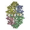
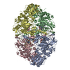
[English] 日本語
Yorodumi
- PDB-5bv2: Crystal structure of E. coli HPII catalase variant -

+
Open data


ID or keywords:

Loading...

-
Basic information

Entry
Database: PDB / ID: 5bv2
TitleCrystal structure of E. coli HPII catalase variant
ComponentsCatalase HPII
KeywordsOXIDOREDUCTASE
Function / homology
Function and homology information


catalase / hyperosmotic response / catalase activity / hydrogen peroxide catabolic process / response to oxidative stress / iron ion binding / DNA damage response / heme binding / identical protein binding / cytoplasm / cytosol
Similarity search - Function
Catalase, four-helical domain / Large catalase, C-terminal domain / C-terminal domain found in long catalases / Catalase, mono-functional, haem-containing, clade 2 / Catalase, mono-functional, haem-containing, clade 2, helical domain / Hemocyanin, N-terminal domain / Catalase HpII, Chain A, domain 1 / Catalase core domain / Catalase haem-binding site / Catalase proximal heme-ligand signature. ...Catalase, four-helical domain / Large catalase, C-terminal domain / C-terminal domain found in long catalases / Catalase, mono-functional, haem-containing, clade 2 / Catalase, mono-functional, haem-containing, clade 2, helical domain / Hemocyanin, N-terminal domain / Catalase HpII, Chain A, domain 1 / Catalase core domain / Catalase haem-binding site / Catalase proximal heme-ligand signature. / Catalase / Catalase active site / Catalase proximal active site signature. / Catalase immune-responsive domain / Catalase-related immune-responsive / Catalase core domain / Catalase, mono-functional, haem-containing / Catalase / catalase family profile. / Catalase superfamily / Class I glutamine amidotransferase (GATase) domain / Class I glutamine amidotransferase-like / Up-down Bundle / Beta Barrel / Rossmann fold / 3-Layer(aba) Sandwich / Mainly Beta / Mainly Alpha / Alpha Beta
Similarity search - Domain/homology
CIS-HEME D HYDROXYCHLORIN GAMMA-SPIROLACTONE / DI(HYDROXYETHYL)ETHER / TRIETHYLENE GLYCOL / Catalase HPII
Similarity search - Component
Biological speciesEscherichia coli (E. coli)
MethodX-RAY DIFFRACTION / SYNCHROTRON / MOLECULAR REPLACEMENT / Resolution: 1.53 Å
AuthorsWang, J. / Lomkalin, I.V.
Funding support United States, 1items
OrganizationGrant numberCountry
National Institutes of Health/National Institute of General Medical Sciences (NIH/NIGMS)GM022778 United States
CitationJournal: To be Published
Title: Influence of weak-intensity data, ordered water molecules, and hydrogen atoms on the refinement of a large protein crystal structure
Authors: Wang, J. / Lomkalin, I.V.
History
DepositionJun 4, 2015Deposition site: RCSB / Processing site: RCSB
Revision 1.0Jun 24, 2015Provider: repository / Type: Initial release
Revision 1.1Feb 14, 2018Group: Advisory / Author supporting evidence ...Advisory / Author supporting evidence / Derived calculations / Source and taxonomy
Category: entity_src_nat / pdbx_audit_support ...entity_src_nat / pdbx_audit_support / pdbx_struct_assembly / pdbx_struct_assembly_gen / pdbx_struct_assembly_prop / pdbx_struct_oper_list / pdbx_validate_close_contact
Item: _entity_src_nat.pdbx_alt_source_flag / _pdbx_audit_support.funding_organization ..._entity_src_nat.pdbx_alt_source_flag / _pdbx_audit_support.funding_organization / _pdbx_audit_support.grant_number / _pdbx_struct_assembly.oligomeric_details / _pdbx_struct_assembly_gen.asym_id_list / _pdbx_struct_assembly_prop.type / _pdbx_struct_assembly_prop.value / _pdbx_struct_oper_list.symmetry_operation
Revision 1.2Dec 25, 2019Group: Author supporting evidence / Category: pdbx_audit_support / Item: _pdbx_audit_support.funding_organization
Revision 1.3Sep 27, 2023Group: Data collection / Database references ...Data collection / Database references / Derived calculations / Refinement description
Category: chem_comp_atom / chem_comp_bond ...chem_comp_atom / chem_comp_bond / database_2 / pdbx_initial_refinement_model / pdbx_struct_conn_angle / struct_conn / struct_conn_type / struct_ncs_dom_lim
Item: _database_2.pdbx_DOI / _database_2.pdbx_database_accession ..._database_2.pdbx_DOI / _database_2.pdbx_database_accession / _pdbx_struct_conn_angle.ptnr1_auth_asym_id / _pdbx_struct_conn_angle.ptnr1_auth_comp_id / _pdbx_struct_conn_angle.ptnr1_auth_seq_id / _pdbx_struct_conn_angle.ptnr1_label_asym_id / _pdbx_struct_conn_angle.ptnr1_label_atom_id / _pdbx_struct_conn_angle.ptnr1_label_comp_id / _pdbx_struct_conn_angle.ptnr1_label_seq_id / _pdbx_struct_conn_angle.ptnr2_auth_asym_id / _pdbx_struct_conn_angle.ptnr2_auth_comp_id / _pdbx_struct_conn_angle.ptnr2_auth_seq_id / _pdbx_struct_conn_angle.ptnr2_label_asym_id / _pdbx_struct_conn_angle.ptnr2_label_atom_id / _pdbx_struct_conn_angle.ptnr2_label_comp_id / _pdbx_struct_conn_angle.ptnr3_auth_asym_id / _pdbx_struct_conn_angle.ptnr3_auth_comp_id / _pdbx_struct_conn_angle.ptnr3_auth_seq_id / _pdbx_struct_conn_angle.ptnr3_label_asym_id / _pdbx_struct_conn_angle.ptnr3_label_atom_id / _pdbx_struct_conn_angle.ptnr3_label_comp_id / _pdbx_struct_conn_angle.value / _struct_conn.conn_type_id / _struct_conn.id / _struct_conn.pdbx_dist_value / _struct_conn.pdbx_leaving_atom_flag / _struct_conn.pdbx_ptnr1_label_alt_id / _struct_conn.pdbx_ptnr2_label_alt_id / _struct_conn.ptnr1_auth_asym_id / _struct_conn.ptnr1_auth_comp_id / _struct_conn.ptnr1_auth_seq_id / _struct_conn.ptnr1_label_asym_id / _struct_conn.ptnr1_label_atom_id / _struct_conn.ptnr1_label_comp_id / _struct_conn.ptnr1_label_seq_id / _struct_conn.ptnr2_auth_asym_id / _struct_conn.ptnr2_auth_comp_id / _struct_conn.ptnr2_auth_seq_id / _struct_conn.ptnr2_label_asym_id / _struct_conn.ptnr2_label_atom_id / _struct_conn.ptnr2_label_comp_id / _struct_conn.ptnr2_label_seq_id / _struct_conn_type.id / _struct_ncs_dom_lim.beg_auth_comp_id / _struct_ncs_dom_lim.beg_label_asym_id / _struct_ncs_dom_lim.beg_label_comp_id / _struct_ncs_dom_lim.beg_label_seq_id / _struct_ncs_dom_lim.end_auth_comp_id / _struct_ncs_dom_lim.end_label_asym_id / _struct_ncs_dom_lim.end_label_comp_id / _struct_ncs_dom_lim.end_label_seq_id

-
Structure visualization

Structure viewerMolecule:
MolmilJmol/JSmol

Downloads & links

-
Assembly

Deposited unit
P: Catalase HPII
Q: Catalase HPII
R: Catalase HPII
S: Catalase HPII
hetero molecules


Theoretical massNumber of molelcules
Total (without water)344,48776
Polymers336,5774
Non-polymers7,91072
Water84,3284681
1


  • Idetical with deposited unit
  • defined by author&software
TypeNameSymmetry operationNumber
identity operation1_555x,y,z1
Buried area79600 Å2
ΔGint-169 kcal/mol
Surface area78550 Å2
MethodPISA
Unit cell
Length a, b, c (Å)161.789, 171.331, 122.484
Angle α, β, γ (deg.)90.00, 121.55, 90.00
Int Tables number5
Space group name H-MC121
Components on special symmetry positions
IDModelComponents
11P-1808-

HOH

21P-1892-

HOH

31P-1906-

HOH

41Q-1537-

HOH

51Q-1941-

HOH

Noncrystallographic symmetry (NCS)NCS domain:
IDEns-IDDetails
11P
21Q
12P
22R
13P
23S
14Q
24R
15Q
25S
16R
26S

NCS domain segments:

Component-ID: _ / Refine code: _

Dom-IDEns-IDBeg auth comp-IDBeg label comp-IDEnd auth comp-IDEnd label comp-IDAuth asym-IDLabel asym-IDAuth seq-IDLabel seq-ID
11ASNASNPROPROPA8 - 7528 - 752
21ASNASNPROPROQB8 - 7528 - 752
12LYSLYSALAALAPA7 - 7537 - 753
22LYSLYSALAALARC7 - 7537 - 753
13LYSLYSALAALAPA7 - 7537 - 753
23LYSLYSALAALASD7 - 7537 - 753
14ASNASNPROPROQB8 - 7528 - 752
24ASNASNPROPRORC8 - 7528 - 752
15ASNASNPROPROQB8 - 7528 - 752
25ASNASNPROPROSD8 - 7528 - 752
16LYSLYSALAALARC7 - 7537 - 753
26LYSLYSALAALASD7 - 7537 - 753

NCS ensembles :
ID
1
2
3
4
5
6

-
Components

-
Protein , 1 types, 4 molecules PQRS

#1: Protein
Catalase HPII / Hydroxyperoxidase II


Mass: 84144.180 Da / Num. of mol.: 4 / Source method: isolated from a natural source / Source: (natural) Escherichia coli (E. coli) / References: UniProt: P21179, catalase

-
Non-polymers , 9 types, 4753 molecules

#2: Chemical
ChemComp-HDD / CIS-HEME D HYDROXYCHLORIN GAMMA-SPIROLACTONE / HEME


Mass: 632.487 Da / Num. of mol.: 4 / Source method: obtained synthetically / Formula: C34H32FeN4O5
#3: Chemical
ChemComp-GOL / GLYCEROL / GLYCERIN / PROPANE-1,2,3-TRIOL


Mass: 92.094 Da / Num. of mol.: 11 / Source method: obtained synthetically / Formula: C3H8O3
#4: Chemical...
ChemComp-EDO / 1,2-ETHANEDIOL / ETHYLENE GLYCOL


Mass: 62.068 Da / Num. of mol.: 39 / Source method: obtained synthetically / Formula: C2H6O2
#5: Chemical ChemComp-PGE / TRIETHYLENE GLYCOL


Mass: 150.173 Da / Num. of mol.: 3 / Source method: obtained synthetically / Formula: C6H14O4
#6: Chemical
ChemComp-PEG / DI(HYDROXYETHYL)ETHER


Mass: 106.120 Da / Num. of mol.: 10 / Source method: obtained synthetically / Formula: C4H10O3
#7: Chemical ChemComp-SO4 / SULFATE ION


Mass: 96.063 Da / Num. of mol.: 2 / Source method: obtained synthetically / Formula: SO4
#8: Chemical ChemComp-MG / MAGNESIUM ION


Mass: 24.305 Da / Num. of mol.: 2 / Source method: obtained synthetically / Formula: Mg
#9: Chemical ChemComp-PG4 / TETRAETHYLENE GLYCOL


Mass: 194.226 Da / Num. of mol.: 1 / Source method: obtained synthetically / Formula: C8H18O5 / Comment: precipitant*YM
#10: Water ChemComp-HOH / water


Mass: 18.015 Da / Num. of mol.: 4681 / Source method: isolated from a natural source / Formula: H2O

-
Experimental details

-
Experiment

ExperimentMethod: X-RAY DIFFRACTION

-
Sample preparation

CrystalDensity Matthews: 2.15 Å3/Da / Density % sol: 42.77 %
Crystal growTemperature: 298 K / Method: vapor diffusion, hanging drop / pH: 7
Details: 2% PEG20000, 15% PEG5000 MME, 0.1 M potassium chloride, 0.1 M manganese acetate, 0.1 M HEPES
PH range: 7

-
Data collection

DiffractionMean temperature: 100 K
Diffraction sourceSource: SYNCHROTRON / Site: NSLS / Beamline: X29A / Wavelength: 1.1 Å
DetectorType: ADSC QUANTUM 315r / Detector: CCD / Date: Mar 1, 2014
RadiationMonochromator: Si(111) / Protocol: SINGLE WAVELENGTH / Monochromatic (M) / Laue (L): M / Scattering type: x-ray
Radiation wavelengthWavelength: 1.1 Å / Relative weight: 1
ReflectionResolution: 1.5→50 Å / Num. all: 451438 / Num. obs: 305824 / % possible obs: 67.7 % / Redundancy: 4.2 % / Rmerge(I) obs: 0.07 / Net I/σ(I): 14.63
Reflection shellHighest resolution: 1.5 Å / CC1/2: 0.46

-
Processing

Software
NameVersionClassification
REFMAC5.8.0073refinement
HKL-3000data reduction
SCALEPACKdata scaling
MOLREPphasing
RefinementMethod to determine structure: MOLECULAR REPLACEMENT
Starting model: PDB entry 4BFL
Resolution: 1.53→50 Å / Cor.coef. Fo:Fc: 0.99 / Cor.coef. Fo:Fc free: 0.975 / SU B: 3.484 / SU ML: 0.05 / Cross valid method: THROUGHOUT / ESU R: 0.152 / ESU R Free: 0.073 / Stereochemistry target values: MAXIMUM LIKELIHOOD / Details: HYDROGENS HAVE BEEN ADDED IN THE RIDING POSITIONS
RfactorNum. reflection% reflectionSelection details
Rfree0.13211 15186 5 %RANDOM
Rwork0.08228 ---
obs0.08475 290638 71.79 %-
Solvent computationIon probe radii: 0.8 Å / Shrinkage radii: 0.8 Å / VDW probe radii: 1.2 Å / Solvent model: MASK
Displacement parametersBiso mean: 18.036 Å2
Baniso -1Baniso -2Baniso -3
1-0.66 Å2-0 Å20.21 Å2
2---0.59 Å2-0 Å2
3----0.19 Å2
Refinement stepCycle: 1 / Resolution: 1.53→50 Å
ProteinNucleic acidLigandSolventTotal
Num. atoms23559 0 535 4669 28763
Refine LS restraints
Refine-IDTypeDev idealDev ideal targetNumber
X-RAY DIFFRACTIONr_bond_refined_d0.0090.01925477
X-RAY DIFFRACTIONr_bond_other_d0.0040.0224026
X-RAY DIFFRACTIONr_angle_refined_deg1.5021.97334728
X-RAY DIFFRACTIONr_angle_other_deg1.107355502
X-RAY DIFFRACTIONr_dihedral_angle_1_deg5.93553217
X-RAY DIFFRACTIONr_dihedral_angle_2_deg35.66423.7771247
X-RAY DIFFRACTIONr_dihedral_angle_3_deg12.809154096
X-RAY DIFFRACTIONr_dihedral_angle_4_deg14.57615196
X-RAY DIFFRACTIONr_chiral_restr0.0790.23658
X-RAY DIFFRACTIONr_gen_planes_refined0.0070.02129102
X-RAY DIFFRACTIONr_gen_planes_other0.0040.025978
X-RAY DIFFRACTIONr_nbd_refined
X-RAY DIFFRACTIONr_nbd_other
X-RAY DIFFRACTIONr_nbtor_refined
X-RAY DIFFRACTIONr_nbtor_other
X-RAY DIFFRACTIONr_xyhbond_nbd_refined
X-RAY DIFFRACTIONr_xyhbond_nbd_other
X-RAY DIFFRACTIONr_metal_ion_refined
X-RAY DIFFRACTIONr_metal_ion_other
X-RAY DIFFRACTIONr_symmetry_vdw_refined
X-RAY DIFFRACTIONr_symmetry_vdw_other
X-RAY DIFFRACTIONr_symmetry_hbond_refined
X-RAY DIFFRACTIONr_symmetry_hbond_other
X-RAY DIFFRACTIONr_symmetry_metal_ion_refined
X-RAY DIFFRACTIONr_symmetry_metal_ion_other
X-RAY DIFFRACTIONr_mcbond_it1.5271.38412199
X-RAY DIFFRACTIONr_mcbond_other1.5271.38412198
X-RAY DIFFRACTIONr_mcangle_it1.9042.08215306
X-RAY DIFFRACTIONr_mcangle_other1.9042.08215307
X-RAY DIFFRACTIONr_scbond_it2.5481.69613274
X-RAY DIFFRACTIONr_scbond_other2.5451.69613270
X-RAY DIFFRACTIONr_scangle_it
X-RAY DIFFRACTIONr_scangle_other2.9712.40419305
X-RAY DIFFRACTIONr_long_range_B_refined6.07915.32734842
X-RAY DIFFRACTIONr_long_range_B_other6.07915.32834843
X-RAY DIFFRACTIONr_rigid_bond_restr1.691349495
X-RAY DIFFRACTIONr_sphericity_free47.4265976
X-RAY DIFFRACTIONr_sphericity_bonded12.211552460
Refine LS restraints NCS

Refine-ID: X-RAY DIFFRACTION / Type: interatomic distance / Weight position: 0.05

Ens-IDDom-IDAuth asym-IDNumberRms dev position (Å)
11P457640.07
12Q457640.07
21P461710.06
22R461710.06
31P462130.06
32S462130.06
41Q460170.07
42R460170.07
51Q461780.07
52S461780.07
61R459610.07
62S459610.07
LS refinement shellResolution: 1.53→1.57 Å / Total num. of bins used: 20
RfactorNum. reflection% reflection
Rfree0.361 33 -
Rwork0.311 795 -
obs--2.63 %

+
About Yorodumi

-
News

-
Feb 9, 2022. New format data for meta-information of EMDB entries

New format data for meta-information of EMDB entries

  • Version 3 of the EMDB header file is now the official format.
  • The previous official version 1.9 will be removed from the archive.

Related info.:EMDB header

External links:wwPDB to switch to version 3 of the EMDB data model

-
Aug 12, 2020. Covid-19 info

Covid-19 info

URL: https://pdbjlvh1.pdbj.org/emnavi/covid19.php

New page: Covid-19 featured information page in EM Navigator.

Related info.:Covid-19 info / Mar 5, 2020. Novel coronavirus structure data

+
Mar 5, 2020. Novel coronavirus structure data

Novel coronavirus structure data

Related info.:Yorodumi Speices / Aug 12, 2020. Covid-19 info

External links:COVID-19 featured content - PDBj / Molecule of the Month (242):Coronavirus Proteases

+
Jan 31, 2019. EMDB accession codes are about to change! (news from PDBe EMDB page)

EMDB accession codes are about to change! (news from PDBe EMDB page)

  • The allocation of 4 digits for EMDB accession codes will soon come to an end. Whilst these codes will remain in use, new EMDB accession codes will include an additional digit and will expand incrementally as the available range of codes is exhausted. The current 4-digit format prefixed with “EMD-” (i.e. EMD-XXXX) will advance to a 5-digit format (i.e. EMD-XXXXX), and so on. It is currently estimated that the 4-digit codes will be depleted around Spring 2019, at which point the 5-digit format will come into force.
  • The EM Navigator/Yorodumi systems omit the EMD- prefix.

Related info.:Q: What is EMD? / ID/Accession-code notation in Yorodumi/EM Navigator

External links:EMDB Accession Codes are Changing Soon! / Contact to PDBj

+
Jul 12, 2017. Major update of PDB

Major update of PDB

  • wwPDB released updated PDB data conforming to the new PDBx/mmCIF dictionary.
  • This is a major update changing the version number from 4 to 5, and with Remediation, in which all the entries are updated.
  • In this update, many items about electron microscopy experimental information are reorganized (e.g. em_software).
  • Now, EM Navigator and Yorodumi are based on the updated data.

External links:wwPDB Remediation / Enriched Model Files Conforming to OneDep Data Standards Now Available in the PDB FTP Archive

-
Yorodumi

Thousand views of thousand structures

  • Yorodumi is a browser for structure data from EMDB, PDB, SASBDB, etc.
  • This page is also the successor to EM Navigator detail page, and also detail information page/front-end page for Omokage search.
  • The word "yorodu" (or yorozu) is an old Japanese word meaning "ten thousand". "mi" (miru) is to see.

Related info.:EMDB / PDB / SASBDB / Comparison of 3 databanks / Yorodumi Search / Aug 31, 2016. New EM Navigator & Yorodumi / Yorodumi Papers / Jmol/JSmol / Function and homology information / Changes in new EM Navigator and Yorodumi

Read more