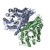
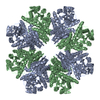
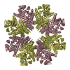
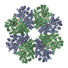
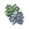
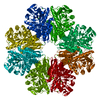
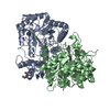
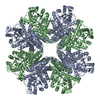
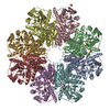
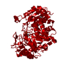
[English] 日本語
Yorodumi
- PDB-5boe: Crystal structure of Staphylococcus aureus enolase in complex with PEP -

+
Open data


ID or keywords:

Loading...

-
Basic information

Entry
Database: PDB / ID: 5boe
TitleCrystal structure of Staphylococcus aureus enolase in complex with PEP
ComponentsEnolase
KeywordsLYASE / enolase / PEP
Function / homology
Function and homology information


phosphopyruvate hydratase / phosphopyruvate hydratase complex / phosphopyruvate hydratase activity / glycolytic process / cell surface / magnesium ion binding / extracellular region
Similarity search - Function
Enolase / Enolase, conserved site / Enolase, C-terminal TIM barrel domain / Enolase, N-terminal / Enolase, C-terminal TIM barrel domain / Enolase, N-terminal domain / Enolase signature. / Enolase, C-terminal TIM barrel domain / Enolase, N-terminal domain / Enolase-like C-terminal domain ...Enolase / Enolase, conserved site / Enolase, C-terminal TIM barrel domain / Enolase, N-terminal / Enolase, C-terminal TIM barrel domain / Enolase, N-terminal domain / Enolase signature. / Enolase, C-terminal TIM barrel domain / Enolase, N-terminal domain / Enolase-like C-terminal domain / Enolase-like, N-terminal domain / Enolase-like, N-terminal / Enolase-like, C-terminal domain superfamily / Enolase-like; domain 1 / TIM Barrel / Alpha-Beta Barrel / 2-Layer Sandwich / Alpha Beta
Similarity search - Domain/homology
PHOSPHOENOLPYRUVATE / Enolase
Similarity search - Component
Biological speciesStaphylococcus aureus (bacteria)
MethodX-RAY DIFFRACTION / SYNCHROTRON / MOLECULAR REPLACEMENT / Resolution: 1.6 Å
AuthorsWang, C.L. / Wu, Y.F. / Han, L. / Wu, M.H. / Zhang, X. / Zang, J.Y.
CitationJournal: Acta Crystallogr.,Sect.D / Year: 2015
Title: Octameric structure of Staphylococcus aureus enolase in complex with phosphoenolpyruvate
Authors: Wu, Y. / Wang, C. / Lin, S. / Wu, M. / Han, L. / Tian, C. / Zhang, X. / Zang, J.
History
DepositionMay 27, 2015Deposition site: RCSB / Processing site: PDBJ
Revision 1.0Dec 9, 2015Provider: repository / Type: Initial release
Revision 1.1Dec 16, 2015Group: Database references
Revision 1.2Nov 8, 2023Group: Data collection / Database references ...Data collection / Database references / Derived calculations / Refinement description
Category: chem_comp_atom / chem_comp_bond ...chem_comp_atom / chem_comp_bond / database_2 / pdbx_initial_refinement_model / pdbx_struct_oper_list / struct_conn / struct_ncs_dom_lim
Item: _database_2.pdbx_DOI / _database_2.pdbx_database_accession ..._database_2.pdbx_DOI / _database_2.pdbx_database_accession / _pdbx_struct_oper_list.symmetry_operation / _struct_conn.pdbx_dist_value / _struct_conn.ptnr1_auth_asym_id / _struct_conn.ptnr1_auth_comp_id / _struct_conn.ptnr1_auth_seq_id / _struct_conn.ptnr1_label_asym_id / _struct_conn.ptnr1_label_atom_id / _struct_conn.ptnr1_label_comp_id / _struct_conn.ptnr1_label_seq_id / _struct_conn.ptnr2_auth_asym_id / _struct_conn.ptnr2_auth_comp_id / _struct_conn.ptnr2_auth_seq_id / _struct_conn.ptnr2_label_asym_id / _struct_conn.ptnr2_label_atom_id / _struct_conn.ptnr2_label_comp_id / _struct_ncs_dom_lim.beg_auth_comp_id / _struct_ncs_dom_lim.beg_label_asym_id / _struct_ncs_dom_lim.beg_label_comp_id / _struct_ncs_dom_lim.beg_label_seq_id / _struct_ncs_dom_lim.end_auth_comp_id / _struct_ncs_dom_lim.end_label_asym_id / _struct_ncs_dom_lim.end_label_comp_id / _struct_ncs_dom_lim.end_label_seq_id

-
Structure visualization

Structure viewerMolecule:
MolmilJmol/JSmol

Downloads & links

-
Assembly

Deposited unit
A: Enolase
B: Enolase
hetero molecules


Theoretical massNumber of molelcules
Total (without water)97,1279
Polymers96,4662
Non-polymers6617
Water11,908661
1
A: Enolase
B: Enolase
hetero molecules

A: Enolase
B: Enolase
hetero molecules

A: Enolase
B: Enolase
hetero molecules

A: Enolase
B: Enolase
hetero molecules


Theoretical massNumber of molelcules
Total (without water)388,50636
Polymers385,8638
Non-polymers2,64428
Water1448
TypeNameSymmetry operationNumber
identity operation1_555x,y,z1
crystal symmetry operation2_555-x,-y,z1
crystal symmetry operation3_555-y,x,z1
crystal symmetry operation4_555y,-x,z1
Buried area31760 Å2
ΔGint-170 kcal/mol
Surface area103830 Å2
MethodPISA
Unit cell
Length a, b, c (Å)145.154, 145.154, 100.512
Angle α, β, γ (deg.)90.00, 90.00, 90.00
Int Tables number79
Space group name H-MI4
Noncrystallographic symmetry (NCS)NCS domain:
IDEns-IDDetails
11A
21B

NCS domain segments:

Component-ID: _ / Ens-ID: 1 / Beg auth comp-ID: MET / Beg label comp-ID: MET / End auth comp-ID: ASP / End label comp-ID: ASP / Refine code: _ / Auth seq-ID: 1 - 433 / Label seq-ID: 1 - 433

Dom-IDAuth asym-IDLabel asym-ID
1AA
2BB

-
Components

#1: Protein Enolase / 2-phospho-D-glycerate hydro-lyase / 2-phosphoglycerate dehydratase / Laminin-binding protein


Mass: 48232.820 Da / Num. of mol.: 2 / Mutation: Q197K
Source method: isolated from a genetically manipulated source
Source: (gene. exp.) Staphylococcus aureus (bacteria) / Gene: eno / Production host: Escherichia coli (E. coli) / References: UniProt: O69174, phosphopyruvate hydratase
#2: Chemical ChemComp-PEP / PHOSPHOENOLPYRUVATE


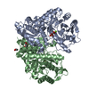
Mass: 168.042 Da / Num. of mol.: 2 / Source method: obtained synthetically / Formula: C3H5O6P
#3: Chemical ChemComp-MG / MAGNESIUM ION


Mass: 24.305 Da / Num. of mol.: 2 / Source method: obtained synthetically / Formula: Mg
#4: Chemical ChemComp-GOL / GLYCEROL / GLYCERIN / PROPANE-1,2,3-TRIOL


Mass: 92.094 Da / Num. of mol.: 3 / Source method: obtained synthetically / Formula: C3H8O3
#5: Water ChemComp-HOH / water


Mass: 18.015 Da / Num. of mol.: 661 / Source method: isolated from a natural source / Formula: H2O

-
Experimental details

-
Experiment

ExperimentMethod: X-RAY DIFFRACTION

-
Sample preparation

CrystalDensity Matthews: 2.74 Å3/Da / Density % sol: 55.18 %
Crystal growTemperature: 289 K / Method: vapor diffusion
Details: 0.2M Ammonium acetate, 0.1M Tris pH 8.5, 25%(w/v) Polyethylene glycol 3,350

-
Data collection

DiffractionMean temperature: 100 K
Diffraction sourceSource: SYNCHROTRON / Site: SSRF / Beamline: BL17U / Wavelength: 0.9792 Å
DetectorType: ADSC QUANTUM 315r / Detector: CCD / Date: Sep 21, 2014
RadiationProtocol: MAD / Monochromatic (M) / Laue (L): M / Scattering type: x-ray
Radiation wavelengthWavelength: 0.9792 Å / Relative weight: 1
ReflectionResolution: 1.6→50 Å / Num. obs: 136548 / % possible obs: 100 % / Redundancy: 8.2 % / Rmerge(I) obs: 0.086 / Net I/σ(I): 40.3
Reflection shellResolution: 1.6→1.63 Å / Redundancy: 8.1 % / Rmerge(I) obs: 0.467 / Mean I/σ(I) obs: 6.8 / % possible all: 100

-
Processing

Software
NameVersionClassification
REFMAC5.8.0073refinement
HKL-2000data reduction
HKL-2000data scaling
PHASERphasing
RefinementMethod to determine structure: MOLECULAR REPLACEMENT
Starting model: 5BOF
Resolution: 1.6→50 Å / Cor.coef. Fo:Fc: 0.969 / Cor.coef. Fo:Fc free: 0.964 / SU B: 1.08 / SU ML: 0.039 / Cross valid method: THROUGHOUT / ESU R: 0.067 / ESU R Free: 0.064 / Stereochemistry target values: MAXIMUM LIKELIHOOD / Details: HYDROGENS HAVE BEEN ADDED IN THE RIDING POSITIONS
RfactorNum. reflection% reflectionSelection details
Rfree0.16356 6858 5 %RANDOM
Rwork0.14848 ---
obs0.14923 129690 99.95 %-
Solvent computationIon probe radii: 0.8 Å / Shrinkage radii: 0.8 Å / VDW probe radii: 1.2 Å / Solvent model: MASK
Displacement parametersBiso mean: 15.677 Å2
Baniso -1Baniso -2Baniso -3
1--0.22 Å20 Å2-0 Å2
2---0.22 Å2-0 Å2
3---0.45 Å2
Refinement stepCycle: LAST / Resolution: 1.6→50 Å
ProteinNucleic acidLigandSolventTotal
Num. atoms6558 0 40 661 7259
Refine LS restraints
Refine-IDTypeDev idealDev ideal targetNumber
X-RAY DIFFRACTIONr_bond_refined_d0.0090.0197029
X-RAY DIFFRACTIONr_bond_other_d0.0050.026582
X-RAY DIFFRACTIONr_angle_refined_deg1.3761.9669574
X-RAY DIFFRACTIONr_angle_other_deg1.047315195
X-RAY DIFFRACTIONr_dihedral_angle_1_deg6.1695941
X-RAY DIFFRACTIONr_dihedral_angle_2_deg39.95825.591313
X-RAY DIFFRACTIONr_dihedral_angle_3_deg11.967151164
X-RAY DIFFRACTIONr_dihedral_angle_4_deg15.1771530
X-RAY DIFFRACTIONr_chiral_restr0.0820.21085
X-RAY DIFFRACTIONr_gen_planes_refined0.0060.028275
X-RAY DIFFRACTIONr_gen_planes_other0.0020.021519
X-RAY DIFFRACTIONr_nbd_refined
X-RAY DIFFRACTIONr_nbd_other
X-RAY DIFFRACTIONr_nbtor_refined
X-RAY DIFFRACTIONr_nbtor_other
X-RAY DIFFRACTIONr_xyhbond_nbd_refined
X-RAY DIFFRACTIONr_xyhbond_nbd_other
X-RAY DIFFRACTIONr_metal_ion_refined
X-RAY DIFFRACTIONr_metal_ion_other
X-RAY DIFFRACTIONr_symmetry_vdw_refined
X-RAY DIFFRACTIONr_symmetry_vdw_other
X-RAY DIFFRACTIONr_symmetry_hbond_refined
X-RAY DIFFRACTIONr_symmetry_hbond_other
X-RAY DIFFRACTIONr_symmetry_metal_ion_refined
X-RAY DIFFRACTIONr_symmetry_metal_ion_other
X-RAY DIFFRACTIONr_mcbond_it0.9451.443662
X-RAY DIFFRACTIONr_mcbond_other0.9361.4393661
X-RAY DIFFRACTIONr_mcangle_it1.5062.1574622
X-RAY DIFFRACTIONr_mcangle_other1.5062.1574623
X-RAY DIFFRACTIONr_scbond_it1.571.6283367
X-RAY DIFFRACTIONr_scbond_other1.5691.6283367
X-RAY DIFFRACTIONr_scangle_it
X-RAY DIFFRACTIONr_scangle_other2.4092.3744948
X-RAY DIFFRACTIONr_long_range_B_refined4.64612.7718767
X-RAY DIFFRACTIONr_long_range_B_other4.52612.5618606
X-RAY DIFFRACTIONr_rigid_bond_restr
X-RAY DIFFRACTIONr_sphericity_free
X-RAY DIFFRACTIONr_sphericity_bonded
Refine LS restraints NCS

Ens-ID: 1 / Number: 27672 / Refine-ID: X-RAY DIFFRACTION / Type: interatomic distance / Rms dev position: 0.08 Å / Weight position: 0.05

Dom-IDAuth asym-ID
1A
2B
LS refinement shellResolution: 1.602→1.643 Å / Total num. of bins used: 20
RfactorNum. reflection% reflection
Rfree0.207 486 -
Rwork0.179 9579 -
obs--99.54 %

+
About Yorodumi

-
News

-
Feb 9, 2022. New format data for meta-information of EMDB entries

New format data for meta-information of EMDB entries

  • Version 3 of the EMDB header file is now the official format.
  • The previous official version 1.9 will be removed from the archive.

Related info.:EMDB header

External links:wwPDB to switch to version 3 of the EMDB data model

-
Aug 12, 2020. Covid-19 info

Covid-19 info

URL: https://pdbjlvh1.pdbj.org/emnavi/covid19.php

New page: Covid-19 featured information page in EM Navigator.

Related info.:Covid-19 info / Mar 5, 2020. Novel coronavirus structure data

+
Mar 5, 2020. Novel coronavirus structure data

Novel coronavirus structure data

Related info.:Yorodumi Speices / Aug 12, 2020. Covid-19 info

External links:COVID-19 featured content - PDBj / Molecule of the Month (242):Coronavirus Proteases

+
Jan 31, 2019. EMDB accession codes are about to change! (news from PDBe EMDB page)

EMDB accession codes are about to change! (news from PDBe EMDB page)

  • The allocation of 4 digits for EMDB accession codes will soon come to an end. Whilst these codes will remain in use, new EMDB accession codes will include an additional digit and will expand incrementally as the available range of codes is exhausted. The current 4-digit format prefixed with “EMD-” (i.e. EMD-XXXX) will advance to a 5-digit format (i.e. EMD-XXXXX), and so on. It is currently estimated that the 4-digit codes will be depleted around Spring 2019, at which point the 5-digit format will come into force.
  • The EM Navigator/Yorodumi systems omit the EMD- prefix.

Related info.:Q: What is EMD? / ID/Accession-code notation in Yorodumi/EM Navigator

External links:EMDB Accession Codes are Changing Soon! / Contact to PDBj

+
Jul 12, 2017. Major update of PDB

Major update of PDB

  • wwPDB released updated PDB data conforming to the new PDBx/mmCIF dictionary.
  • This is a major update changing the version number from 4 to 5, and with Remediation, in which all the entries are updated.
  • In this update, many items about electron microscopy experimental information are reorganized (e.g. em_software).
  • Now, EM Navigator and Yorodumi are based on the updated data.

External links:wwPDB Remediation / Enriched Model Files Conforming to OneDep Data Standards Now Available in the PDB FTP Archive

-
Yorodumi

Thousand views of thousand structures

  • Yorodumi is a browser for structure data from EMDB, PDB, SASBDB, etc.
  • This page is also the successor to EM Navigator detail page, and also detail information page/front-end page for Omokage search.
  • The word "yorodu" (or yorozu) is an old Japanese word meaning "ten thousand". "mi" (miru) is to see.

Related info.:EMDB / PDB / SASBDB / Comparison of 3 databanks / Yorodumi Search / Aug 31, 2016. New EM Navigator & Yorodumi / Yorodumi Papers / Jmol/JSmol / Function and homology information / Changes in new EM Navigator and Yorodumi

Read more