 Movie
Movie Controller

[English] 日本語
 Yorodumi
- PDB-5b52: Crystal structure of the N-terminal domain of H-NS family protein TurB -
+ Open data

- Basic information
| Entry | Database: PDB / ID: 5b52 | ||||||
|---|---|---|---|---|---|---|---|
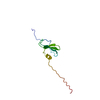
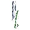
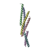
| Title | Crystal structure of the N-terminal domain of H-NS family protein TurB | ||||||
 Components Components | H-NS family protein MvaT | ||||||
 Keywords Keywords | TRANSCRIPTION / transcriptional regulator / nucleoid associated protein | ||||||
| Function / homology | MvaT, DNA-binding domain / Bacterial xenogeneic silencer MvaT, C-terminal domain / H-NS family protein MvaT Function and homology information Function and homology information | ||||||
| Biological species |  Pseudomonas putida (bacteria) Pseudomonas putida (bacteria) | ||||||
| Method |  X-RAY DIFFRACTION / SYNCHROTRON / SAD / Resolution: 2.3 Å X-RAY DIFFRACTION / SYNCHROTRON / SAD / Resolution: 2.3 Å | ||||||
 Authors Authors | Suzuki-Minakuchi, C. / Nojiri, H. | ||||||
| Funding support |  Japan, 1items Japan, 1items
| ||||||
 Citation Citation | Journal: Febs Lett. / Year: 2016 Title: Structural similarities and differences in H-NS family proteins revealed by the N-terminal structure of TurB in Pseudomonas putida KT2440 Authors: Suzuki-Minakuchi, C. / Kawazuma, K. / Matsuzawa, J. / Vasileva, D. / Fujimoto, Z. / Terada, T. / Okada, K. / Nojiri, H. | ||||||
| History |
|
- Structure visualization
| Structure viewer | Molecule: MolmilJmol/JSmol |
|---|
- Downloads & links
-Download
| PDBx/mmCIF format | 5b52.cif.gz | 35.7 KB | Display | PDBx/mmCIF format |
|---|---|---|---|---|
| PDB format | pdb5b52.ent.gz | 25 KB | Display | PDB format |
| PDBx/mmJSON format | 5b52.json.gz | Tree view | PDBx/mmJSON format | |
| Others |  Other downloads Other downloads |
-Validation report
| Arichive directory | https://data.pdbj.org/pub/pdb/validation_reports/b5/5b52ftp://data.pdbj.org/pub/pdb/validation_reports/b5/5b52 | HTTPS FTP |
|---|
-Related structure data
| Similar structure data |
|---|


-Links
 PDBj
PDBj- Assembly
| Deposited unit | 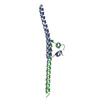
| ||||||||
|---|---|---|---|---|---|---|---|---|---|
| 1 |
| ||||||||
| Unit cell |
| ||||||||
| Components on special symmetry positions |
|
-Components
| #1: Protein | Mass: 8004.292 Da / Num. of mol.: 2 / Fragment: UNP residues 1-61 / Mutation: R8A Source method: isolated from a genetically manipulated source Source: (gene. exp.) Pseudomonas putida (strain KT2440) (bacteria)Strain: KT2440 / Gene: PP_3765 / Production host: #2: Water | ChemComp-HOH / |  Mass: 18.015 Da / Num. of mol.: 48 / Source method: isolated from a natural source / Formula: H2O Mass: 18.015 Da / Num. of mol.: 48 / Source method: isolated from a natural source / Formula: H2O |
|---|
-Experimental details
-Experiment
| Experiment | Method: X-RAY DIFFRACTION / Number of used crystals: 1 |
|---|
- Sample preparation
| Crystal | Density Matthews: 2.59 Å3/Da / Density % sol: 52.45 % |
|---|---|
| Crystal grow | Temperature: 293 K / Method: vapor diffusion, hanging drop / Details: ammonium acetate, BIS-TRIS propane / PH range: 6.9-7.0 |
-Data collection
| Diffraction |
| |||||||||||||||||||||
|---|---|---|---|---|---|---|---|---|---|---|---|---|---|---|---|---|---|---|---|---|---|---|
| Diffraction source |
| |||||||||||||||||||||
| Detector |
| |||||||||||||||||||||
| Radiation |
| |||||||||||||||||||||
| Radiation wavelength |
| |||||||||||||||||||||
| Reflection | Entry-ID: 5B52
| |||||||||||||||||||||
| Reflection shell |
|
- Processing
| Software |
| ||||||||||||||||||||||||||||||||||||||||||||||||||||||||||||||||||||||||||||||||||||||||||||||||||||||||||||||||||||||||||||||||||||||||||||||||||||||||||||||||||||||||||||||||||||||
|---|---|---|---|---|---|---|---|---|---|---|---|---|---|---|---|---|---|---|---|---|---|---|---|---|---|---|---|---|---|---|---|---|---|---|---|---|---|---|---|---|---|---|---|---|---|---|---|---|---|---|---|---|---|---|---|---|---|---|---|---|---|---|---|---|---|---|---|---|---|---|---|---|---|---|---|---|---|---|---|---|---|---|---|---|---|---|---|---|---|---|---|---|---|---|---|---|---|---|---|---|---|---|---|---|---|---|---|---|---|---|---|---|---|---|---|---|---|---|---|---|---|---|---|---|---|---|---|---|---|---|---|---|---|---|---|---|---|---|---|---|---|---|---|---|---|---|---|---|---|---|---|---|---|---|---|---|---|---|---|---|---|---|---|---|---|---|---|---|---|---|---|---|---|---|---|---|---|---|---|---|---|---|---|
| Refinement | Method to determine structure: SAD / Resolution: 2.3→52.03 Å / Cor.coef. Fo:Fc: 0.927 / Cor.coef. Fo:Fc free: 0.881 / SU B: 8.811 / SU ML: 0.2 / Cross valid method: THROUGHOUT / ESU R: 0.309 / ESU R Free: 0.257 / Stereochemistry target values: MAXIMUM LIKELIHOOD / Details: HYDROGENS HAVE BEEN ADDED IN THE RIDING POSITIONS
| ||||||||||||||||||||||||||||||||||||||||||||||||||||||||||||||||||||||||||||||||||||||||||||||||||||||||||||||||||||||||||||||||||||||||||||||||||||||||||||||||||||||||||||||||||||||
| Solvent computation | Ion probe radii: 0.8 Å / Shrinkage radii: 0.8 Å / VDW probe radii: 1.2 Å / Solvent model: MASK | ||||||||||||||||||||||||||||||||||||||||||||||||||||||||||||||||||||||||||||||||||||||||||||||||||||||||||||||||||||||||||||||||||||||||||||||||||||||||||||||||||||||||||||||||||||||
| Displacement parameters | Biso mean: 43.261 Å2
| ||||||||||||||||||||||||||||||||||||||||||||||||||||||||||||||||||||||||||||||||||||||||||||||||||||||||||||||||||||||||||||||||||||||||||||||||||||||||||||||||||||||||||||||||||||||
| Refinement step | Cycle: LAST / Resolution: 2.3→52.03 Å
| ||||||||||||||||||||||||||||||||||||||||||||||||||||||||||||||||||||||||||||||||||||||||||||||||||||||||||||||||||||||||||||||||||||||||||||||||||||||||||||||||||||||||||||||||||||||
| Refine LS restraints |
|