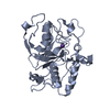
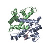
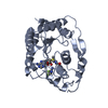
 Movie
Movie Controller


+ Open data

- Basic information
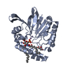
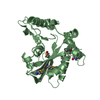
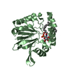
| Entry | Database: PDB / ID: 5b4c | ||||||
|---|---|---|---|---|---|---|---|
| Title | Crystal structure of H10N mutant of LpxH with manganese | ||||||
 Components Components | UDP-2,3-diacylglucosamine hydrolase | ||||||
 Keywords Keywords | HYDROLASE / LpxH / Lipid A / Lipid X / UDP-2 / 3-diacylglucosamine | ||||||
| Function / homology |  Function and homology information Function and homology informationUDP-2,3-diacylglucosamine diphosphatase / UDP-2,3-diacylglucosamine hydrolase activity / glycolipid binding / extrinsic component of plasma membrane / lipid A biosynthetic process / manganese ion binding / cytoplasm Similarity search - Function | ||||||
| Biological species |  Pseudomonas aeruginosa PAO1 (bacteria) Pseudomonas aeruginosa PAO1 (bacteria) | ||||||
| Method |  X-RAY DIFFRACTION / SYNCHROTRON / MOLECULAR REPLACEMENT / Resolution: 1.96 Å X-RAY DIFFRACTION / SYNCHROTRON / MOLECULAR REPLACEMENT / Resolution: 1.96 Å | ||||||
 Authors Authors | Okada, C. / Wakabayashi, H. / Yao, M. / Tanaka, I. | ||||||
 Citation Citation | Journal: Sci Rep / Year: 2016 Title: Crystal structures of the UDP-diacylglucosamine pyrophosphohydrase LpxH from Pseudomonas aeruginosa Authors: Okada, C. / Wakabayashi, H. / Kobayashi, M. / Shinoda, A. / Tanaka, I. / Yao, M. | ||||||
| History |
|
- Structure visualization
| Structure viewer | Molecule: MolmilJmol/JSmol |
|---|
- Downloads & links
-Download
| PDBx/mmCIF format | 5b4c.cif.gz | 205.8 KB | Display | PDBx/mmCIF format |
|---|---|---|---|---|
| PDB format | pdb5b4c.ent.gz | 163.5 KB | Display | PDB format |
| PDBx/mmJSON format | 5b4c.json.gz | Tree view | PDBx/mmJSON format | |
| Others |  Other downloads Other downloads |
-Validation report
| Summary document | 5b4c_validation.pdf.gz | 446 KB | Display | wwPDB validaton report |
|---|---|---|---|---|
| Full document | 5b4c_full_validation.pdf.gz | 446.7 KB | Display | |
| Data in XML | 5b4c_validation.xml.gz | 23.1 KB | Display | |
| Data in CIF | 5b4c_validation.cif.gz | 34.4 KB | Display | |
| Arichive directory | https://data.pdbj.org/pub/pdb/validation_reports/b4/5b4cftp://data.pdbj.org/pub/pdb/validation_reports/b4/5b4c | HTTPS FTP |
-Related structure data
| Related structure data |  5b49SC  5b4aC 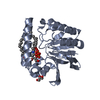 5b4bC  5b4dC S: Starting model for refinement C: citing same article ( |
|---|---|
| Similar structure data |

-Links
 PDBj
PDBj
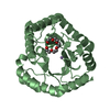
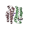
- Assembly
| Deposited unit | 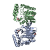
| ||||||||
|---|---|---|---|---|---|---|---|---|---|
| 1 | 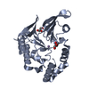
| ||||||||
| 2 | 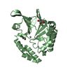
| ||||||||
| Unit cell |
|
-Components
| #1: Protein | Mass: 28646.891 Da / Num. of mol.: 2 / Mutation: H10N Source method: isolated from a genetically manipulated source Source: (gene. exp.) Pseudomonas aeruginosa PAO1 (bacteria) / Strain: PAO1 / Gene: lpxH, PA1792 / Production host: References: UniProt: Q9I2V0, UDP-2,3-diacylglucosamine diphosphatase #2: Chemical |   Mass: 54.938 Da / Num. of mol.: 2 / Source method: obtained synthetically / Formula: Mn Mass: 54.938 Da / Num. of mol.: 2 / Source method: obtained synthetically / Formula: Mn#3: Chemical | ChemComp-GOL /   Mass: 92.094 Da / Num. of mol.: 4 / Source method: obtained synthetically / Formula: C3H8O3 Mass: 92.094 Da / Num. of mol.: 4 / Source method: obtained synthetically / Formula: C3H8O3#4: Water | ChemComp-HOH / |  Mass: 18.015 Da / Num. of mol.: 442 / Source method: isolated from a natural source / Formula: H2O Mass: 18.015 Da / Num. of mol.: 442 / Source method: isolated from a natural source / Formula: H2O |
|---|
-Experimental details
-Experiment
| Experiment | Method: X-RAY DIFFRACTION / Number of used crystals: 1 |
|---|
- Sample preparation
| Crystal | Density Matthews: 2.61 Å3/Da / Density % sol: 52.79 % |
|---|---|
| Crystal grow | Temperature: 293 K / Method: vapor diffusion, sitting drop / pH: 6.7 / Details: 0.2M Proline, 0.1M MES pH 6.7, 12.5% PEG 3350 |
-Data collection
| Diffraction | Mean temperature: 100 K |
|---|---|
| Diffraction source | Source: SYNCHROTRON / Site: SPring-8  / Beamline: BL32XU / Wavelength: 1 Å / Beamline: BL32XU / Wavelength: 1 Å |
| Detector | Type: RAYONIX MX225HE / Detector: CCD / Date: Nov 11, 2011 |
| Radiation | Protocol: SINGLE WAVELENGTH / Monochromatic (M) / Laue (L): M / Scattering type: x-ray |
| Radiation wavelength | Wavelength: 1 Å / Relative weight: 1 |
| Reflection | Resolution: 1.95→50 Å / Num. obs: 43726 / % possible obs: 99.7 % / Redundancy: 3.6 % / Rsym value: 0.078 / Net I/σ(I): 17.7 |
| Reflection shell | Resolution: 1.95→2.02 Å / Redundancy: 3.6 % / Rsym value: 0.451 / % possible all: 99.8 |
- Processing
| Software |
| ||||||||||||||||||||||||||||||||||||||||||||||||||||||||||||||||||||||||||||||||||||||||||||||||||||||||||||||||||||||||||||||||||||||||||||||||||||||||||||||||||||||||||||||||||||||
|---|---|---|---|---|---|---|---|---|---|---|---|---|---|---|---|---|---|---|---|---|---|---|---|---|---|---|---|---|---|---|---|---|---|---|---|---|---|---|---|---|---|---|---|---|---|---|---|---|---|---|---|---|---|---|---|---|---|---|---|---|---|---|---|---|---|---|---|---|---|---|---|---|---|---|---|---|---|---|---|---|---|---|---|---|---|---|---|---|---|---|---|---|---|---|---|---|---|---|---|---|---|---|---|---|---|---|---|---|---|---|---|---|---|---|---|---|---|---|---|---|---|---|---|---|---|---|---|---|---|---|---|---|---|---|---|---|---|---|---|---|---|---|---|---|---|---|---|---|---|---|---|---|---|---|---|---|---|---|---|---|---|---|---|---|---|---|---|---|---|---|---|---|---|---|---|---|---|---|---|---|---|---|---|
| Refinement | Method to determine structure: MOLECULAR REPLACEMENT Starting model: 5B49 Resolution: 1.96→49.15 Å / Cor.coef. Fo:Fc: 0.962 / Cor.coef. Fo:Fc free: 0.942 / SU B: 6.369 / SU ML: 0.093 / Cross valid method: THROUGHOUT / ESU R: 0.141 / ESU R Free: 0.136 / Details: HYDROGENS HAVE BEEN ADDED IN THE RIDING POSITIONS
| ||||||||||||||||||||||||||||||||||||||||||||||||||||||||||||||||||||||||||||||||||||||||||||||||||||||||||||||||||||||||||||||||||||||||||||||||||||||||||||||||||||||||||||||||||||||
| Solvent computation | Ion probe radii: 0.8 Å / Shrinkage radii: 0.8 Å / VDW probe radii: 1.2 Å | ||||||||||||||||||||||||||||||||||||||||||||||||||||||||||||||||||||||||||||||||||||||||||||||||||||||||||||||||||||||||||||||||||||||||||||||||||||||||||||||||||||||||||||||||||||||
| Displacement parameters | Biso mean: 32.591 Å2
| ||||||||||||||||||||||||||||||||||||||||||||||||||||||||||||||||||||||||||||||||||||||||||||||||||||||||||||||||||||||||||||||||||||||||||||||||||||||||||||||||||||||||||||||||||||||
| Refinement step | Cycle: LAST / Resolution: 1.96→49.15 Å
| ||||||||||||||||||||||||||||||||||||||||||||||||||||||||||||||||||||||||||||||||||||||||||||||||||||||||||||||||||||||||||||||||||||||||||||||||||||||||||||||||||||||||||||||||||||||
| Refine LS restraints |
|