 Movie
Movie Controller

[English] 日本語
 Yorodumi
- PDB-5awe: Crystal structure of a hypothetical protein, TTHA0829 from Thermu... -
+ Open data

- Basic information
| Entry | Database: PDB / ID: 5awe | ||||||
|---|---|---|---|---|---|---|---|
| Title | Crystal structure of a hypothetical protein, TTHA0829 from Thermus thermophilus HB8, composed of cystathionine-beta-synthase (CBS) and aspartate-kinase chorismate-mutase tyrA (ACT) domains | ||||||
 Components Components | Putative acetoin utilization protein, acetoin dehydrogenase | ||||||
 Keywords Keywords | STRUCTURAL GENOMICS / UNKNOWN FUNCTION / hypothetical protein / Thermus thermophilus HB8 / CBS domain / ACT domain / RIKEN Structural Genomics/Proteomics Initiative / RSGI | ||||||
| Function / homology |  Function and homology information Function and homology informationTTHA0829-like ACT domain / : / CBS-domain / CBS-domain / Domain in cystathionine beta-synthase and other proteins. / CBS domain superfamily / CBS domain / CBS domain / CBS domain profile. / Roll / Alpha Beta Similarity search - Domain/homology | ||||||
| Biological species |   Thermus thermophilus (bacteria) Thermus thermophilus (bacteria) | ||||||
| Method |  X-RAY DIFFRACTION / SYNCHROTRON / SAD / Resolution: 2.45 Å X-RAY DIFFRACTION / SYNCHROTRON / SAD / Resolution: 2.45 Å | ||||||
 Authors Authors | Nakabayashi, M. / Shibata, N. / Kanagawa, M. / Nakagawa, N. / Kuramitsu, S. / Higuchi, Y. | ||||||
 Citation Citation | Journal: Extremophiles / Year: 2016 Title: Crystal structure of a hypothetical protein, TTHA0829 from Thermus thermophilus HB8, composed of cystathionine-beta-synthase (CBS) and aspartate-kinase chorismate-mutase tyrA (ACT) domains. Authors: Nakabayashi, M. / Shibata, N. / Ishido-Nakai, E. / Kanagawa, M. / Iio, Y. / Komori, H. / Ueda, Y. / Nakagawa, N. / Kuramitsu, S. / Higuchi, Y. | ||||||
| History |
|
- Structure visualization
| Structure viewer | Molecule: MolmilJmol/JSmol |
|---|
- Downloads & links
-Download
| PDBx/mmCIF format | 5awe.cif.gz | 54 KB | Display | PDBx/mmCIF format |
|---|---|---|---|---|
| PDB format | pdb5awe.ent.gz | 38.4 KB | Display | PDB format |
| PDBx/mmJSON format | 5awe.json.gz | Tree view | PDBx/mmJSON format | |
| Others |  Other downloads Other downloads |
-Validation report
| Arichive directory | https://data.pdbj.org/pub/pdb/validation_reports/aw/5aweftp://data.pdbj.org/pub/pdb/validation_reports/aw/5awe | HTTPS FTP |
|---|
-Related structure data
| Related structure data | |
|---|---|
| Similar structure data |


-Links
 PDBj
PDBj
- Assembly
| Deposited unit | 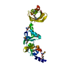
| ||||||||
|---|---|---|---|---|---|---|---|---|---|
| 1 | 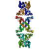
| ||||||||
| Unit cell |
|
-Components
| #1: Protein | Mass: 23251.953 Da / Num. of mol.: 1 Source method: isolated from a genetically manipulated source Source: (gene. exp.) Thermus thermophilus (bacteria) / Strain: HB8 / ATCC 27634 / DSM 579 / Gene: TTHA0829 / Plasmid: pET11a / Production host: Escherichia coli / Strain (production host): B834 (DE3) / References: UniProt: Q5SK23 |
|---|---|
| #2: Water | ChemComp-HOH /  Mass: 18.015 Da / Num. of mol.: 53 / Source method: isolated from a natural source / Formula: H2O Mass: 18.015 Da / Num. of mol.: 53 / Source method: isolated from a natural source / Formula: H2O |
| Has protein modification | Y |
-Experimental details
-Experiment
| Experiment | Method: X-RAY DIFFRACTION |
|---|
- Sample preparation
| Crystal | Density Matthews: 2.3 Å3/Da / Density % sol: 46.44 % / Description: THE FILE CONTAINS FRIEDEL PAIRS. |
|---|---|
| Crystal grow | Temperature: 293 K / Method: vapor diffusion, sitting drop / pH: 7.5 / Details: HEPES, magnesium chloride, PEG 400 |
-Data collection
| Diffraction | Mean temperature: 100 K |
|---|---|
| Diffraction source | Source: SYNCHROTRON / Site: SPring-8  / Beamline: BL26B1 / Wavelength: 0.97911 Å / Beamline: BL26B1 / Wavelength: 0.97911 Å |
| Detector | Type: RIGAKU JUPITER 210 / Detector: CCD / Date: Jun 25, 2004 |
| Radiation | Protocol: SINGLE WAVELENGTH / Monochromatic (M) / Laue (L): M / Scattering type: x-ray |
| Radiation wavelength | Wavelength: 0.97911 Å / Relative weight: 1 |
| Reflection | Resolution: 2.45→40.51 Å / Num. obs: 8426 / % possible obs: 99.6 % / Redundancy: 16.4 % / Biso Wilson estimate: 29.5 Å2 / Rmerge(I) obs: 0.104 / Net I/σ(I): 31.7 |
| Reflection shell | Resolution: 2.45→2.54 Å / Redundancy: 14.1 % / Rmerge(I) obs: 0.305 / Mean I/σ(I) obs: 4.6 / % possible all: 98.2 |
- Processing
| Software |
| ||||||||||||||||||||||||||||||||||||||||||||||||||||||||||||
|---|---|---|---|---|---|---|---|---|---|---|---|---|---|---|---|---|---|---|---|---|---|---|---|---|---|---|---|---|---|---|---|---|---|---|---|---|---|---|---|---|---|---|---|---|---|---|---|---|---|---|---|---|---|---|---|---|---|---|---|---|---|
| Refinement | Method to determine structure: SAD / Resolution: 2.45→40.51 Å / Rfactor Rfree error: 0.007 / Data cutoff high absF: 246220.86 / Data cutoff low absF: 0 / Isotropic thermal model: RESTRAINED / Cross valid method: THROUGHOUT / σ(F): 0 Details: THE FILE CONTAINS FRIEDEL PAIRS. BULK SOLVENT MODEL USED.
| ||||||||||||||||||||||||||||||||||||||||||||||||||||||||||||
| Solvent computation | Solvent model: FLAT MODEL / Bsol: 48.9401 Å2 / ksol: 0.38 e/Å3 | ||||||||||||||||||||||||||||||||||||||||||||||||||||||||||||
| Displacement parameters | Biso mean: 35.8 Å2
| ||||||||||||||||||||||||||||||||||||||||||||||||||||||||||||
| Refine analyze |
| ||||||||||||||||||||||||||||||||||||||||||||||||||||||||||||
| Refinement step | Cycle: 1 / Resolution: 2.45→40.51 Å /
| ||||||||||||||||||||||||||||||||||||||||||||||||||||||||||||
| Refine LS restraints |
| ||||||||||||||||||||||||||||||||||||||||||||||||||||||||||||
| LS refinement shell | Resolution: 2.45→2.54 Å / Rfactor Rfree error: 0.043 / Total num. of bins used: 10
| ||||||||||||||||||||||||||||||||||||||||||||||||||||||||||||
| Xplor file |
|
