[English] 日本語
Yorodumi
- PDB-5anw: MTH1 in complex with compound 24 -

+
Open data


ID or keywords:

Loading...

-
Basic information

Entry
Database: PDB / ID: 5anw
TitleMTH1 in complex with compound 24
Components7,8-DIHYDRO-8-OXOGUANINE TRIPHOSPHATASE
KeywordsHYDROLASE / NUDT1 / NUDIX HYDROLASE / INHIBITOR
Function / homology
Function and homology information


2-hydroxy-ATP hydrolase activity / 2-hydroxy-dATP hydrolase activity / N6-methyl-(d)ATP hydrolase activity / O6-methyl-dGTP hydrolase activity / 2-hydroxy-dATP diphosphatase / dATP diphosphatase activity / ATP diphosphatase activity / 8-oxo-7,8-dihydrodeoxyguanosine triphosphate pyrophosphatase activity / hydrolase activity, acting on acid anhydrides, in phosphorus-containing anhydrides / 8-oxo-7,8-dihydroguanosine triphosphate pyrophosphatase activity ...2-hydroxy-ATP hydrolase activity / 2-hydroxy-dATP hydrolase activity / N6-methyl-(d)ATP hydrolase activity / O6-methyl-dGTP hydrolase activity / 2-hydroxy-dATP diphosphatase / dATP diphosphatase activity / ATP diphosphatase activity / 8-oxo-7,8-dihydrodeoxyguanosine triphosphate pyrophosphatase activity / hydrolase activity, acting on acid anhydrides, in phosphorus-containing anhydrides / 8-oxo-7,8-dihydroguanosine triphosphate pyrophosphatase activity / DNA protection / Phosphate bond hydrolysis by NUDT proteins / purine nucleoside catabolic process / snoRNA binding / Hydrolases; Acting on acid anhydrides; In phosphorus-containing anhydrides / response to cadmium ion / acrosomal vesicle / male gonad development / nuclear membrane / response to oxidative stress / mitochondrial matrix / DNA repair / mitochondrion / extracellular space / nucleus / metal ion binding / cytoplasm / cytosol
Similarity search - Function
Oxidized purine nucleoside triphosphate / NUDIX hydrolase / NUDIX hydrolase, conserved site / Nudix box signature. / Nucleoside Triphosphate Pyrophosphohydrolase / Nucleoside Triphosphate Pyrophosphohydrolase / NUDIX domain / Nudix hydrolase domain profile. / NUDIX hydrolase domain / NUDIX hydrolase-like domain superfamily ...Oxidized purine nucleoside triphosphate / NUDIX hydrolase / NUDIX hydrolase, conserved site / Nudix box signature. / Nucleoside Triphosphate Pyrophosphohydrolase / Nucleoside Triphosphate Pyrophosphohydrolase / NUDIX domain / Nudix hydrolase domain profile. / NUDIX hydrolase domain / NUDIX hydrolase-like domain superfamily / Alpha-Beta Complex / Alpha Beta
Similarity search - Domain/homology
Chem-9CQ / Oxidized purine nucleoside triphosphate hydrolase
Similarity search - Component
Biological speciesHOMO SAPIENS (human)
MethodX-RAY DIFFRACTION / SYNCHROTRON / MOLECULAR REPLACEMENT / Resolution: 1.37 Å
AuthorsRead, J.A. / Breed, J.
CitationJournal: J.Med.Chem. / Year: 2016
Title: Potent and Selective Inhibitors of Mth1 Probe its Role in Cancer Cell Survival.
Authors: Kettle, J.G. / Alwan, H. / Bista, M. / Breed, J. / Davies, N.L. / Eckersley, K. / Fillery, S. / Foote, K.M. / Goodwin, L. / Jones, D.R. / Kack, H. / Lau, A. / Nissink, J.W. / Read, J. / ...Authors: Kettle, J.G. / Alwan, H. / Bista, M. / Breed, J. / Davies, N.L. / Eckersley, K. / Fillery, S. / Foote, K.M. / Goodwin, L. / Jones, D.R. / Kack, H. / Lau, A. / Nissink, J.W. / Read, J. / Scott, J.S. / Taylor, B. / Walker, G. / Wissler, L. / Wylot, M.
History
DepositionSep 8, 2015Deposition site: PDBE / Processing site: PDBE
Revision 1.0Mar 2, 2016Provider: repository / Type: Initial release
Revision 1.1Mar 16, 2016Group: Atomic model
Revision 1.2Apr 6, 2016Group: Database references
Revision 1.3May 8, 2024Group: Data collection / Database references ...Data collection / Database references / Derived calculations / Other
Category: chem_comp_atom / chem_comp_bond ...chem_comp_atom / chem_comp_bond / database_2 / pdbx_database_status / struct_site
Item: _database_2.pdbx_DOI / _database_2.pdbx_database_accession ..._database_2.pdbx_DOI / _database_2.pdbx_database_accession / _pdbx_database_status.status_code_sf / _struct_site.pdbx_auth_asym_id / _struct_site.pdbx_auth_comp_id / _struct_site.pdbx_auth_seq_id

-
Structure visualization

Structure viewerMolecule:
MolmilJmol/JSmol

Downloads & links

-
Assembly

Deposited unit
A: 7,8-DIHYDRO-8-OXOGUANINE TRIPHOSPHATASE
hetero molecules


Theoretical massNumber of molelcules
Total (without water)18,4062
Polymers18,1001
Non-polymers3061
Water1,31573
1


  • Idetical with deposited unit
  • defined by software
TypeNameSymmetry operationNumber
identity operation1_555x,y,z1
MethodPISA
Unit cell
Length a, b, c (Å)61.130, 66.070, 35.980
Angle α, β, γ (deg.)90.00, 90.00, 90.00
Int Tables number18
Space group name H-MP21212
Components on special symmetry positions
IDModelComponents
11A-2029-

HOH

21A-2068-

HOH

-
Components

#1: Protein 7,8-DIHYDRO-8-OXOGUANINE TRIPHOSPHATASE / MTH1 / 2-HYDROXY-DATP DIPHOSPHATASE / 8-OXO-DGTPASE / NUCLEOSIDE DIPHOSPHATE-LINKED MOIETY X MOTIF ...MTH1 / 2-HYDROXY-DATP DIPHOSPHATASE / 8-OXO-DGTPASE / NUCLEOSIDE DIPHOSPHATE-LINKED MOIETY X MOTIF 1 / NUDIX MOTIF 1


Mass: 18099.590 Da / Num. of mol.: 1
Source method: isolated from a genetically manipulated source
Source: (gene. exp.) HOMO SAPIENS (human) / Production host: ESCHERICHIA COLI (E. coli) / Strain (production host): BL21(DE3) / Variant (production host): GOLD
References: UniProt: P36639, 8-oxo-dGTP diphosphatase, 2-hydroxy-dATP diphosphatase
#2: Chemical ChemComp-9CQ / 2-[4-(2-AMINOQUINAZOLIN-4-YL)PHENYL]-N,N-DIMETHYL-ACETAMIDE


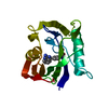
Mass: 306.362 Da / Num. of mol.: 1 / Source method: obtained synthetically / Formula: C18H18N4O
#3: Water ChemComp-HOH / water


Mass: 18.015 Da / Num. of mol.: 73 / Source method: isolated from a natural source / Formula: H2O

-
Experimental details

-
Experiment

ExperimentMethod: X-RAY DIFFRACTION / Number of used crystals: 1

-
Sample preparation

CrystalDensity Matthews: 2.01 Å3/Da / Density % sol: 38.72 % / Description: NONE
Crystal growpH: 4.5 / Details: pH 4.5

-
Data collection

DiffractionMean temperature: 100 K
Diffraction sourceSource: SYNCHROTRON / Site: Diamond / Beamline: I03 / Wavelength: 0.97625
DetectorType: DECTRIS PILATUS / Detector: PIXEL / Date: Jul 4, 2015 / Details: MIRRORS
RadiationProtocol: SINGLE WAVELENGTH / Monochromatic (M) / Laue (L): M / Scattering type: x-ray
Radiation wavelengthWavelength: 0.97625 Å / Relative weight: 1
ReflectionResolution: 1.37→61.1 Å / Num. obs: 25048 / % possible obs: 80 % / Observed criterion σ(I): 2 / Redundancy: 5 % / Biso Wilson estimate: 13.27 Å2 / Rmerge(I) obs: 0.05 / Net I/σ(I): 24.6
Reflection shellResolution: 1.37→1.41 Å / Redundancy: 1.1 % / Rmerge(I) obs: 0.4 / Mean I/σ(I) obs: 4.8 / % possible all: 17

-
Processing

Software
NameVersionClassification
BUSTER2.11.6refinement
MOSFLMdata reduction
Aimlessdata scaling
AMoREphasing
RefinementMethod to determine structure: MOLECULAR REPLACEMENT / Resolution: 1.37→15.86 Å / Cor.coef. Fo:Fc: 0.9419 / Cor.coef. Fo:Fc free: 0.9352 / SU R Cruickshank DPI: 0.077 / Cross valid method: THROUGHOUT / σ(F): 0 / SU R Blow DPI: 0.078 / SU Rfree Blow DPI: 0.078 / SU Rfree Cruickshank DPI: 0.077
Details: IDEAL-DIST CONTACT TERM CONTACT SETUP. ALL ATOMS HAVE CCP4 ATOM TYPE FROM LIBRARY
RfactorNum. reflection% reflectionSelection details
Rfree0.2253 1218 4.88 %RANDOM
Rwork0.1989 ---
obs0.2003 24983 79.71 %-
Displacement parametersBiso mean: 14.98 Å2
Baniso -1Baniso -2Baniso -3
1-0.2861 Å20 Å20 Å2
2--0.5173 Å20 Å2
3----0.8033 Å2
Refine analyzeLuzzati coordinate error obs: 0.198 Å
Refinement stepCycle: LAST / Resolution: 1.37→15.86 Å
ProteinNucleic acidLigandSolventTotal
Num. atoms1210 0 23 73 1306
Refine LS restraints
Refine-IDTypeDev idealNumberRestraint functionWeight
X-RAY DIFFRACTIONt_bond_d0.0081320HARMONIC2
X-RAY DIFFRACTIONt_angle_deg0.971804HARMONIC2
X-RAY DIFFRACTIONt_dihedral_angle_d438SINUSOIDAL2
X-RAY DIFFRACTIONt_incorr_chiral_ct
X-RAY DIFFRACTIONt_pseud_angle
X-RAY DIFFRACTIONt_trig_c_planes31HARMONIC2
X-RAY DIFFRACTIONt_gen_planes198HARMONIC5
X-RAY DIFFRACTIONt_it1320HARMONIC20
X-RAY DIFFRACTIONt_nbd
X-RAY DIFFRACTIONt_omega_torsion4.09
X-RAY DIFFRACTIONt_other_torsion14.84
X-RAY DIFFRACTIONt_improper_torsion
X-RAY DIFFRACTIONt_chiral_improper_torsion160SEMIHARMONIC5
X-RAY DIFFRACTIONt_sum_occupancies
X-RAY DIFFRACTIONt_utility_distance
X-RAY DIFFRACTIONt_utility_angle
X-RAY DIFFRACTIONt_utility_torsion
X-RAY DIFFRACTIONt_ideal_dist_contact1494SEMIHARMONIC4
LS refinement shellResolution: 1.37→1.43 Å / Total num. of bins used: 13
RfactorNum. reflection% reflection
Rfree0.3417 32 4.23 %
Rwork0.3707 724 -
all0.3693 756 -
obs--21.77 %
Refinement TLS params.Method: refined / Origin x: 14.2828 Å / Origin y: 19.754 Å / Origin z: 9.2814 Å
111213212223313233
T-0.0126 Å20.0081 Å2-0.0089 Å2-0.0017 Å20.0027 Å2---0.0076 Å2
L0.8015 °20.464 °2-0.0704 °2-1.2662 °2-0.1466 °2--0.6698 °2
S0.041 Å °0.0228 Å °-0.037 Å °0.0549 Å °0.003 Å °0.0204 Å °0.003 Å °-0.0375 Å °-0.044 Å °
Refinement TLS groupSelection details: { A|3 - A|156 }

+
About Yorodumi

-
News

-
Feb 9, 2022. New format data for meta-information of EMDB entries

New format data for meta-information of EMDB entries

  • Version 3 of the EMDB header file is now the official format.
  • The previous official version 1.9 will be removed from the archive.

Related info.:EMDB header

External links:wwPDB to switch to version 3 of the EMDB data model

-
Aug 12, 2020. Covid-19 info

Covid-19 info

URL: https://pdbjlvh1.pdbj.org/emnavi/covid19.php

New page: Covid-19 featured information page in EM Navigator.

Related info.:Covid-19 info / Mar 5, 2020. Novel coronavirus structure data

+
Mar 5, 2020. Novel coronavirus structure data

Novel coronavirus structure data

Related info.:Yorodumi Speices / Aug 12, 2020. Covid-19 info

External links:COVID-19 featured content - PDBj / Molecule of the Month (242):Coronavirus Proteases

+
Jan 31, 2019. EMDB accession codes are about to change! (news from PDBe EMDB page)

EMDB accession codes are about to change! (news from PDBe EMDB page)

  • The allocation of 4 digits for EMDB accession codes will soon come to an end. Whilst these codes will remain in use, new EMDB accession codes will include an additional digit and will expand incrementally as the available range of codes is exhausted. The current 4-digit format prefixed with “EMD-” (i.e. EMD-XXXX) will advance to a 5-digit format (i.e. EMD-XXXXX), and so on. It is currently estimated that the 4-digit codes will be depleted around Spring 2019, at which point the 5-digit format will come into force.
  • The EM Navigator/Yorodumi systems omit the EMD- prefix.

Related info.:Q: What is EMD? / ID/Accession-code notation in Yorodumi/EM Navigator

External links:EMDB Accession Codes are Changing Soon! / Contact to PDBj

+
Jul 12, 2017. Major update of PDB

Major update of PDB

  • wwPDB released updated PDB data conforming to the new PDBx/mmCIF dictionary.
  • This is a major update changing the version number from 4 to 5, and with Remediation, in which all the entries are updated.
  • In this update, many items about electron microscopy experimental information are reorganized (e.g. em_software).
  • Now, EM Navigator and Yorodumi are based on the updated data.

External links:wwPDB Remediation / Enriched Model Files Conforming to OneDep Data Standards Now Available in the PDB FTP Archive

-
Yorodumi

Thousand views of thousand structures

  • Yorodumi is a browser for structure data from EMDB, PDB, SASBDB, etc.
  • This page is also the successor to EM Navigator detail page, and also detail information page/front-end page for Omokage search.
  • The word "yorodu" (or yorozu) is an old Japanese word meaning "ten thousand". "mi" (miru) is to see.

Related info.:EMDB / PDB / SASBDB / Comparison of 3 databanks / Yorodumi Search / Aug 31, 2016. New EM Navigator & Yorodumi / Yorodumi Papers / Jmol/JSmol / Function and homology information / Changes in new EM Navigator and Yorodumi

Read more