 Movie
Movie Controller

[English] 日本語
 Yorodumi
- PDB-5an4: Crystal structure of the human 8-oxoguanine glycosylase (OGG1) pr... -
+ Open data

- Basic information
| Entry | Database: PDB / ID: 5an4 | ||||||
|---|---|---|---|---|---|---|---|
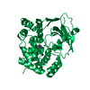
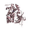
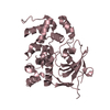
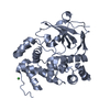
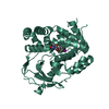
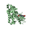
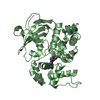
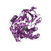
| Title | Crystal structure of the human 8-oxoguanine glycosylase (OGG1) processed with the CrystalDirect automated mounting and cryo-cooling technology | ||||||
 Components Components | N-GLYCOSYLASE/DNA LYASE | ||||||
 Keywords Keywords | HYDROLASE / ALLERGEN / PYR/PYL/RCAR / BET V 1 / FLAVONOIDS / AUTOMATED CRYSTAL HARVESTING / AUTOMATED CRYO-COOLING / CRYSTALDIRECT | ||||||
| Function / homology |  Function and homology information Function and homology informationDefective OGG1 Substrate Binding / Defective OGG1 Substrate Processing / Defective OGG1 Localization / depurination / negative regulation of double-strand break repair via single-strand annealing / oxidized purine nucleobase lesion DNA N-glycosylase activity / base-excision repair, AP site formation / depyrimidination / 8-oxo-7,8-dihydroguanine DNA N-glycosylase activity / Displacement of DNA glycosylase by APEX1 ...Defective OGG1 Substrate Binding / Defective OGG1 Substrate Processing / Defective OGG1 Localization / depurination / negative regulation of double-strand break repair via single-strand annealing / oxidized purine nucleobase lesion DNA N-glycosylase activity / base-excision repair, AP site formation / depyrimidination / 8-oxo-7,8-dihydroguanine DNA N-glycosylase activity / Displacement of DNA glycosylase by APEX1 / positive regulation of gene expression via chromosomal CpG island demethylation / Hydrolases; Glycosylases; Hydrolysing N-glycosyl compounds / oxidized purine DNA binding / APEX1-Independent Resolution of AP Sites via the Single Nucleotide Replacement Pathway / Recognition and association of DNA glycosylase with site containing an affected purine / Cleavage of the damaged purine / Recognition and association of DNA glycosylase with site containing an affected pyrimidine / Cleavage of the damaged pyrimidine / class I DNA-(apurinic or apyrimidinic site) endonuclease activity / DNA-(apurinic or apyrimidinic site) lyase / nucleotide-excision repair / cellular response to reactive oxygen species / response to radiation / base-excision repair / nuclear matrix / response to oxidative stress / endonuclease activity / microtubule binding / damaged DNA binding / nuclear speck / RNA polymerase II cis-regulatory region sequence-specific DNA binding / mitochondrial matrix / DNA damage response / regulation of DNA-templated transcription / enzyme binding / positive regulation of transcription by RNA polymerase II / protein-containing complex / mitochondrion / DNA binding / nucleoplasm / nucleus / cytosol Similarity search - Function | ||||||
| Biological species |  HOMO SAPIENS (human) HOMO SAPIENS (human) | ||||||
| Method |  X-RAY DIFFRACTION / SYNCHROTRON / OTHER / Resolution: 1.6 Å X-RAY DIFFRACTION / SYNCHROTRON / OTHER / Resolution: 1.6 Å | ||||||
 Authors Authors | Zander, U. / Ytre-Arne, M. / Dalhus, B. / Hoffmann, G. / Cornaciu, I. / Cipriani, F. / Marquez, J.A. | ||||||
 Citation Citation | Journal: Acta Crystallogr.,Sect.D / Year: 2016 Title: Automated Harvesting and Processing of Protein Crystals Through Laser Photoablation. Authors: Zander, U. / Hoffmann, G. / Cornaciu, I. / Marquette, J.-P. / Papp, G. / Landret, C. / Seroul, G. / Sinoir, J. / Roewer, M. / Felisaz, F. / Rodriguez-Puente, S. / Mariaule, V. / Murphy, P. / ...Authors: Zander, U. / Hoffmann, G. / Cornaciu, I. / Marquette, J.-P. / Papp, G. / Landret, C. / Seroul, G. / Sinoir, J. / Roewer, M. / Felisaz, F. / Rodriguez-Puente, S. / Mariaule, V. / Murphy, P. / Mathieu, M. / Cipriani, F. / Marquez, J.A. | ||||||
| History |
|
- Structure visualization
| Structure viewer | Molecule: MolmilJmol/JSmol |
|---|
- Downloads & links
-Download
| PDBx/mmCIF format | 5an4.cif.gz | 76.4 KB | Display | PDBx/mmCIF format |
|---|---|---|---|---|
| PDB format | pdb5an4.ent.gz | 56.8 KB | Display | PDB format |
| PDBx/mmJSON format | 5an4.json.gz | Tree view | PDBx/mmJSON format | |
| Others |  Other downloads Other downloads |
-Validation report
| Arichive directory | https://data.pdbj.org/pub/pdb/validation_reports/an/5an4ftp://data.pdbj.org/pub/pdb/validation_reports/an/5an4 | HTTPS FTP |
|---|
-Related structure data
| Related structure data |  5amwC  5amxC  5amzC 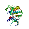 5andC 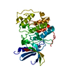 5aneC 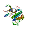 5angC  5aniC  5anjC  5ankC  5anlC 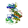 5anoC  5dwpC  5ebhC  5an2 C: citing same article ( |
|---|---|
| Similar structure data |


-Links
 PDBj
PDBj


- Assembly
| Deposited unit | 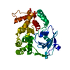
| ||||||||
|---|---|---|---|---|---|---|---|---|---|
| 1 |
| ||||||||
| Unit cell |
|
-Components
| #1: Protein | Mass: 35089.742 Da / Num. of mol.: 1 / Fragment: UNP RESIDUES 12-323 Source method: isolated from a genetically manipulated source Source: (gene. exp.) HOMO SAPIENS (human) / Production host:  References: UniProt: O15527, DNA-formamidopyrimidine glycosylase, DNA-(apurinic or apyrimidinic site) lyase | ||
|---|---|---|---|
| #2: Chemical |   Mass: 96.063 Da / Num. of mol.: 2 / Source method: obtained synthetically / Formula: SO4 Mass: 96.063 Da / Num. of mol.: 2 / Source method: obtained synthetically / Formula: SO4#3: Water | ChemComp-HOH / |  Mass: 18.015 Da / Num. of mol.: 157 / Source method: isolated from a natural source / Formula: H2O Mass: 18.015 Da / Num. of mol.: 157 / Source method: isolated from a natural source / Formula: H2O |
-Experimental details
-Experiment
| Experiment | Method: X-RAY DIFFRACTION |
|---|
- Sample preparation
| Crystal | Density Matthews: 2.19 Å3/Da / Density % sol: 43.8 % / Description: NONE |
|---|---|
| Crystal grow | Details: 0.1 M SODIUM CITRATE, PH=5.5, 0.2 M AMMONIUM SULFATE, 24 % PEG 3350 |
-Data collection
| Diffraction | Mean temperature: 100 K |
|---|---|
| Diffraction source | Source: SYNCHROTRON / Site: ESRF  / Beamline: ID29 / Wavelength: 0.9793 / Beamline: ID29 / Wavelength: 0.9793 |
| Detector | Type: DECTRIS PILATUS 6M / Detector: PIXEL |
| Radiation | Protocol: SINGLE WAVELENGTH / Monochromatic (M) / Laue (L): M / Scattering type: x-ray |
| Radiation wavelength | Wavelength: 0.9793 Å / Relative weight: 1 |
| Reflection | Resolution: 1.6→27 Å / Num. obs: 40235 / % possible obs: 99.9 % / Observed criterion σ(I): 3 / Redundancy: 7 % / Rmerge(I) obs: 0.08 |
- Processing
| Software | Name: REFMAC / Version: 5.8.0123 / Classification: refinement | ||||||||||||||||||||||||||||||||||||||||||||||||||||||||||||||||||||||||||||||||||||||||||||||||||||||||||||||||||||||||||||||||||||||||||||||||||||||||||||||||||||||||||||||||||||||
|---|---|---|---|---|---|---|---|---|---|---|---|---|---|---|---|---|---|---|---|---|---|---|---|---|---|---|---|---|---|---|---|---|---|---|---|---|---|---|---|---|---|---|---|---|---|---|---|---|---|---|---|---|---|---|---|---|---|---|---|---|---|---|---|---|---|---|---|---|---|---|---|---|---|---|---|---|---|---|---|---|---|---|---|---|---|---|---|---|---|---|---|---|---|---|---|---|---|---|---|---|---|---|---|---|---|---|---|---|---|---|---|---|---|---|---|---|---|---|---|---|---|---|---|---|---|---|---|---|---|---|---|---|---|---|---|---|---|---|---|---|---|---|---|---|---|---|---|---|---|---|---|---|---|---|---|---|---|---|---|---|---|---|---|---|---|---|---|---|---|---|---|---|---|---|---|---|---|---|---|---|---|---|---|
| Refinement | Method to determine structure: OTHER Starting model: NONE Resolution: 1.6→26.52 Å / Cor.coef. Fo:Fc: 0.958 / Cor.coef. Fo:Fc free: 0.944 / SU B: 1.84 / SU ML: 0.064 / Cross valid method: THROUGHOUT / ESU R: 0.091 / ESU R Free: 0.091 / Stereochemistry target values: MAXIMUM LIKELIHOOD Details: HYDROGENS HAVE BEEN ADDED IN THE RIDING POSITIONS. U VALUES, REFINED INDIVIDUALLY
| ||||||||||||||||||||||||||||||||||||||||||||||||||||||||||||||||||||||||||||||||||||||||||||||||||||||||||||||||||||||||||||||||||||||||||||||||||||||||||||||||||||||||||||||||||||||
| Solvent computation | Ion probe radii: 0.8 Å / Shrinkage radii: 0.8 Å / VDW probe radii: 1.2 Å / Solvent model: MASK | ||||||||||||||||||||||||||||||||||||||||||||||||||||||||||||||||||||||||||||||||||||||||||||||||||||||||||||||||||||||||||||||||||||||||||||||||||||||||||||||||||||||||||||||||||||||
| Displacement parameters | Biso mean: 18.633 Å2
| ||||||||||||||||||||||||||||||||||||||||||||||||||||||||||||||||||||||||||||||||||||||||||||||||||||||||||||||||||||||||||||||||||||||||||||||||||||||||||||||||||||||||||||||||||||||
| Refinement step | Cycle: LAST / Resolution: 1.6→26.52 Å
| ||||||||||||||||||||||||||||||||||||||||||||||||||||||||||||||||||||||||||||||||||||||||||||||||||||||||||||||||||||||||||||||||||||||||||||||||||||||||||||||||||||||||||||||||||||||
| Refine LS restraints |
|