 Movie
Movie Controller


+ Open data

- Basic information
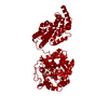
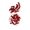
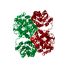
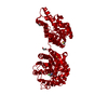
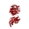
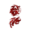
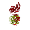
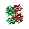
| Entry | Database: PDB / ID: 5alo | ||||||
|---|---|---|---|---|---|---|---|
| Title | ligand complex structure of soluble epoxide hydrolase | ||||||
 Components Components | BIFUNCTIONAL EPOXIDE HYDROLASE 2 | ||||||
 Keywords Keywords | HYDROLASE | ||||||
| Function / homology |  Function and homology information Function and homology informationlipid-phosphate phosphatase / 10-hydroxy-9-(phosphonooxy)octadecanoate phosphatase activity / stilbene catabolic process / Biosynthesis of maresins / epoxide metabolic process / phospholipid dephosphorylation / lipid phosphatase activity / soluble epoxide hydrolase / Synthesis of epoxy (EET) and dihydroxyeicosatrienoic acids (DHET) / lysophosphatidic acid phosphatase activity ...lipid-phosphate phosphatase / 10-hydroxy-9-(phosphonooxy)octadecanoate phosphatase activity / stilbene catabolic process / Biosynthesis of maresins / epoxide metabolic process / phospholipid dephosphorylation / lipid phosphatase activity / soluble epoxide hydrolase / Synthesis of epoxy (EET) and dihydroxyeicosatrienoic acids (DHET) / lysophosphatidic acid phosphatase activity / epoxide hydrolase activity / dephosphorylation / regulation of cholesterol metabolic process / peroxisomal matrix / phosphatase activity / toxic substance binding / cholesterol homeostasis / Peroxisomal protein import / regulation of cell growth / response to toxic substance / peroxisome / positive regulation of gene expression / magnesium ion binding / protein homodimerization activity / extracellular exosome / cytosol Similarity search - Function | ||||||
| Biological species |  HOMO SAPIENS (human) HOMO SAPIENS (human) | ||||||
| Method |  X-RAY DIFFRACTION / MOLECULAR REPLACEMENT / Resolution: 2 Å X-RAY DIFFRACTION / MOLECULAR REPLACEMENT / Resolution: 2 Å | ||||||
 Authors Authors | Oster, L. / Tapani, S. / Xue, Y. / Kack, H. | ||||||
 Citation Citation | Journal: Drug Discov Today / Year: 2015 Title: Successful Generation of Structural Information for Fragment-Based Drug Discovery. Authors: Oster, L. / Tapani, S. / Xue, Y. / Kack, H. | ||||||
| History |
|
- Structure visualization
| Structure viewer | Molecule: MolmilJmol/JSmol |
|---|
- Downloads & links
-Download
| PDBx/mmCIF format | 5alo.cif.gz | 131.9 KB | Display | PDBx/mmCIF format |
|---|---|---|---|---|
| PDB format | pdb5alo.ent.gz | 102.4 KB | Display | PDB format |
| PDBx/mmJSON format | 5alo.json.gz | Tree view | PDBx/mmJSON format | |
| Others |  Other downloads Other downloads |
-Validation report
| Arichive directory | https://data.pdbj.org/pub/pdb/validation_reports/al/5aloftp://data.pdbj.org/pub/pdb/validation_reports/al/5alo | HTTPS FTP |
|---|
-Related structure data
| Related structure data | 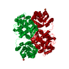 5ahxSC 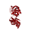 5ai0C 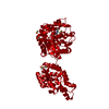 5ai4C 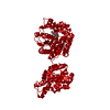 5ai5C 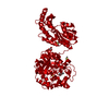 5ai6C 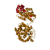 5ai8C 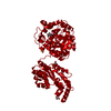 5ai9C  5aiaC 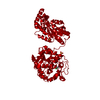 5aibC 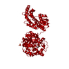 5aicC 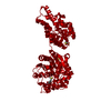 5ak3C 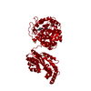 5ak4C 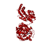 5ak5C 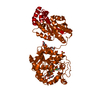 5ak6C  5akeC 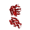 5akgC  5akhC 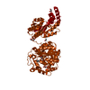 5akiC 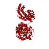 5akjC 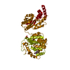 5akkC 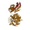 5aklC 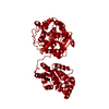 5akxC 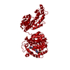 5akyC 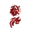 5akzC 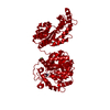 5aldC 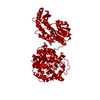 5aleC 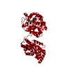 5alfC  5algC 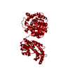 5alhC 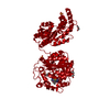 5aliC  5aljC 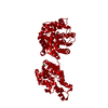 5alkC 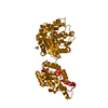 5allC  5almC 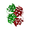 5alnC  5alpC 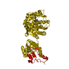 5alqC 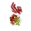 5alrC 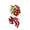 5alsC 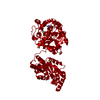 5altC 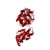 5aluC 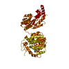 5alvC 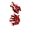 5alwC 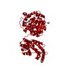 5alxC 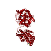 5alyC 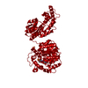 5alzC 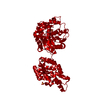 5am0C 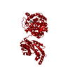 5am1C 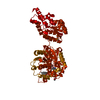 5am2C 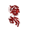 5am3C 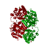 5am4C  5am5C C: citing same article ( S: Starting model for refinement |
|---|---|
| Similar structure data |


-Links
 PDBj
PDBj


- Assembly
| Deposited unit | 
| ||||||||
|---|---|---|---|---|---|---|---|---|---|
| 1 | 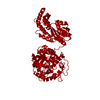
| ||||||||
| Unit cell |
|
-Components
| #1: Protein | Mass: 62002.711 Da / Num. of mol.: 1 Source method: isolated from a genetically manipulated source Source: (gene. exp.) HOMO SAPIENS (human) / Cell line (production host): SF9 / Production host:   SPODOPTERA FRUGIPERDA (fall armyworm) SPODOPTERA FRUGIPERDA (fall armyworm)References: UniProt: P34913, soluble epoxide hydrolase, lipid-phosphate phosphatase | ||||
|---|---|---|---|---|---|
| #2: Chemical |   Mass: 78.133 Da / Num. of mol.: 3 / Source method: obtained synthetically / Formula: C2H6OS / Comment: DMSO, precipitant*YM Mass: 78.133 Da / Num. of mol.: 3 / Source method: obtained synthetically / Formula: C2H6OS / Comment: DMSO, precipitant*YM#3: Chemical | ChemComp-A0J / | 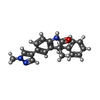  Mass: 317.384 Da / Num. of mol.: 1 / Source method: obtained synthetically / Formula: C20H19N3O Mass: 317.384 Da / Num. of mol.: 1 / Source method: obtained synthetically / Formula: C20H19N3O#4: Water | ChemComp-HOH / |  Mass: 18.015 Da / Num. of mol.: 453 / Source method: isolated from a natural source / Formula: H2O Mass: 18.015 Da / Num. of mol.: 453 / Source method: isolated from a natural source / Formula: H2O |
-Experimental details
-Experiment
| Experiment | Method: X-RAY DIFFRACTION |
|---|
- Sample preparation
| Crystal | Density Matthews: 2.47 Å3/Da / Density % sol: 50.12 % / Description: NONE |
|---|
-Data collection
| Diffraction | Mean temperature: 100 K |
|---|---|
| Diffraction source | Source: ROTATING ANODE / Type: RIGAKU FR-E+ / Wavelength: 1.5418 |
| Detector | Type: RIGAKU CCD-A200-CU / Detector: CCD / Details: MIRRORS |
| Radiation | Protocol: SINGLE WAVELENGTH / Monochromatic (M) / Laue (L): M / Scattering type: x-ray |
| Radiation wavelength | Wavelength: 1.5418 Å / Relative weight: 1 |
| Reflection | Resolution: 2→245.64 Å / Num. obs: 43151 / % possible obs: 99.2 % / Observed criterion σ(I): 0 / Redundancy: 11.5 % / Biso Wilson estimate: 33.15 Å2 / Rmerge(I) obs: 0.09 / Net I/σ(I): 26.8 |
| Reflection shell | Resolution: 2→2.1 Å / Redundancy: 11.2 % / Rmerge(I) obs: 1 / Mean I/σ(I) obs: 2.6 / % possible all: 98.2 |
- Processing
| Software |
| ||||||||||||||||||||||||||||||||||||||||||||||||||||||||||||||||||||||||||||||||||||||||||||||||||||||||||||||||||
|---|---|---|---|---|---|---|---|---|---|---|---|---|---|---|---|---|---|---|---|---|---|---|---|---|---|---|---|---|---|---|---|---|---|---|---|---|---|---|---|---|---|---|---|---|---|---|---|---|---|---|---|---|---|---|---|---|---|---|---|---|---|---|---|---|---|---|---|---|---|---|---|---|---|---|---|---|---|---|---|---|---|---|---|---|---|---|---|---|---|---|---|---|---|---|---|---|---|---|---|---|---|---|---|---|---|---|---|---|---|---|---|---|---|---|---|
| Refinement | Method to determine structure: MOLECULAR REPLACEMENT Starting model: PDB ENTRY 5AHX Resolution: 2→80.42 Å / Cor.coef. Fo:Fc: 0.9457 / Cor.coef. Fo:Fc free: 0.9151 / SU R Cruickshank DPI: 0.178 / Cross valid method: THROUGHOUT / σ(F): 0 / SU R Blow DPI: 0.196 / SU Rfree Blow DPI: 0.167 / SU Rfree Cruickshank DPI: 0.16
| ||||||||||||||||||||||||||||||||||||||||||||||||||||||||||||||||||||||||||||||||||||||||||||||||||||||||||||||||||
| Displacement parameters | Biso mean: 40.55 Å2
| ||||||||||||||||||||||||||||||||||||||||||||||||||||||||||||||||||||||||||||||||||||||||||||||||||||||||||||||||||
| Refine analyze | Luzzati coordinate error obs: 0.237 Å | ||||||||||||||||||||||||||||||||||||||||||||||||||||||||||||||||||||||||||||||||||||||||||||||||||||||||||||||||||
| Refinement step | Cycle: LAST / Resolution: 2→80.42 Å
| ||||||||||||||||||||||||||||||||||||||||||||||||||||||||||||||||||||||||||||||||||||||||||||||||||||||||||||||||||
| Refine LS restraints |
| ||||||||||||||||||||||||||||||||||||||||||||||||||||||||||||||||||||||||||||||||||||||||||||||||||||||||||||||||||
| LS refinement shell | Resolution: 2→2.05 Å / Total num. of bins used: 20
|
