 Movie
Movie Controller

[English] 日本語
 Yorodumi
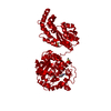
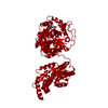
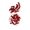
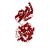
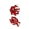
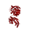
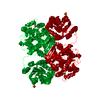
- PDB-4y2j: Structure of soluble epoxide hydrolase in complex with N-[(1-meth... -
+ Open data

- Basic information
| Entry | Database: PDB / ID: 4y2j | ||||||
|---|---|---|---|---|---|---|---|
| Title | Structure of soluble epoxide hydrolase in complex with N-[(1-methyl-1H-pyrazol-3-yl)methyl]-2-phenylethanamine | ||||||
 Components Components | Bifunctional epoxide hydrolase 2 | ||||||
 Keywords Keywords | OXIDOREDUCTASE/OXIDOREDUCTASE INHIBITOR / hydrolase / OXIDOREDUCTASE-OXIDOREDUCTASE INHIBITOR complex | ||||||
| Function / homology |  Function and homology information Function and homology informationlipid-phosphate phosphatase / 10-hydroxy-9-(phosphonooxy)octadecanoate phosphatase activity / stilbene catabolic process / Biosynthesis of maresins / epoxide metabolic process / phospholipid dephosphorylation / lipid phosphatase activity / soluble epoxide hydrolase / Synthesis of epoxy (EET) and dihydroxyeicosatrienoic acids (DHET) / lysophosphatidic acid phosphatase activity ...lipid-phosphate phosphatase / 10-hydroxy-9-(phosphonooxy)octadecanoate phosphatase activity / stilbene catabolic process / Biosynthesis of maresins / epoxide metabolic process / phospholipid dephosphorylation / lipid phosphatase activity / soluble epoxide hydrolase / Synthesis of epoxy (EET) and dihydroxyeicosatrienoic acids (DHET) / lysophosphatidic acid phosphatase activity / epoxide hydrolase activity / dephosphorylation / regulation of cholesterol metabolic process / peroxisomal matrix / phosphatase activity / toxic substance binding / cholesterol homeostasis / Peroxisomal protein import / regulation of cell growth / response to toxic substance / peroxisome / positive regulation of gene expression / magnesium ion binding / protein homodimerization activity / extracellular exosome / cytosol Similarity search - Function | ||||||
| Biological species |  Homo sapiens (human) Homo sapiens (human) | ||||||
| Method |  X-RAY DIFFRACTION / SYNCHROTRON / Resolution: 2.15 Å X-RAY DIFFRACTION / SYNCHROTRON / Resolution: 2.15 Å | ||||||
 Authors Authors | Amano, Y. / Yamaguchi, T. | ||||||
 Citation Citation | Journal: Bioorg.Med.Chem. / Year: 2015 Title: Identification of N-ethylmethylamine as a novel scaffold for inhibitors of soluble epoxide hydrolase by crystallographic fragment screening Authors: Amano, Y. / Tanabe, E. / Yamaguchi, T. | ||||||
| History |
|
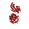
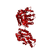
- Structure visualization
| Structure viewer | Molecule: MolmilJmol/JSmol |
|---|
- Downloads & links
-Download
| PDBx/mmCIF format | 4y2j.cif.gz | 123.9 KB | Display | PDBx/mmCIF format |
|---|---|---|---|---|
| PDB format | pdb4y2j.ent.gz | 95.3 KB | Display | PDB format |
| PDBx/mmJSON format | 4y2j.json.gz | Tree view | PDBx/mmJSON format | |
| Others |  Other downloads Other downloads |
-Validation report
| Arichive directory | https://data.pdbj.org/pub/pdb/validation_reports/y2/4y2jftp://data.pdbj.org/pub/pdb/validation_reports/y2/4y2j | HTTPS FTP |
|---|
-Related structure data
| Related structure data | 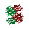 4y2pC 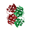 4y2qC  4y2rC 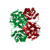 4y2sC 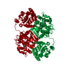 4y2tC 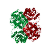 4y2uC 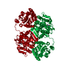 4y2vC 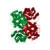 4y2xC 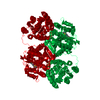 4y2yC C: citing same article ( |
|---|---|
| Similar structure data |


-Links
 PDBj
PDBj


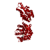
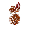
- Assembly
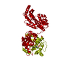
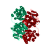
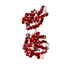
| Deposited unit | 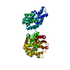
| ||||||||
|---|---|---|---|---|---|---|---|---|---|
| 1 | 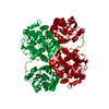
| ||||||||
| Unit cell |
|
-Components
| #1: Protein | Mass: 63514.512 Da / Num. of mol.: 1 / Fragment: UNP residues 1-555 Source method: isolated from a genetically manipulated source Source: (gene. exp.) Homo sapiens (human) / Gene: EPHX2 / Production host:  References: UniProt: P34913, soluble epoxide hydrolase, lipid-phosphate phosphatase |
|---|---|
| #2: Chemical | ChemComp-MG /   Mass: 24.305 Da / Num. of mol.: 1 / Source method: obtained synthetically / Formula: Mg Mass: 24.305 Da / Num. of mol.: 1 / Source method: obtained synthetically / Formula: Mg |
| #3: Chemical | ChemComp-49G / 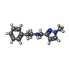  Mass: 215.294 Da / Num. of mol.: 1 / Source method: obtained synthetically / Formula: C13H17N3 Mass: 215.294 Da / Num. of mol.: 1 / Source method: obtained synthetically / Formula: C13H17N3 |
| #4: Water | ChemComp-HOH /  Mass: 18.015 Da / Num. of mol.: 104 / Source method: isolated from a natural source / Formula: H2O Mass: 18.015 Da / Num. of mol.: 104 / Source method: isolated from a natural source / Formula: H2O |
-Experimental details
-Experiment
| Experiment | Method: X-RAY DIFFRACTION |
|---|
- Sample preparation
| Crystal | Density Matthews: 2.39 Å3/Da / Density % sol: 48.44 % |
|---|---|
| Crystal grow | Temperature: 298 K / Method: vapor diffusion, sitting drop / pH: 7.5 Details: Potassium phosphate, Ammonium dihydrogen phosphate, PEG3350 |
-Data collection
| Diffraction | Mean temperature: 90 K |
|---|---|
| Diffraction source | Source: SYNCHROTRON / Site: Photon Factory  / Beamline: AR-NW12A / Wavelength: 1 Å / Beamline: AR-NW12A / Wavelength: 1 Å |
| Detector | Type: ADSC QUANTUM 210 / Detector: CCD / Date: May 31, 2007 |
| Radiation | Protocol: SINGLE WAVELENGTH / Monochromatic (M) / Laue (L): M / Scattering type: x-ray |
| Radiation wavelength | Wavelength: 1 Å / Relative weight: 1 |
| Reflection | Resolution: 2.15→80.33 Å / Num. obs: 34505 / % possible obs: 99.1 % / Redundancy: 7.2 % / Net I/σ(I): 25.4 |
| Reflection shell | Resolution: 2.15→2.23 Å / Redundancy: 7.3 % / Rmerge(I) obs: 0.372 / Mean I/σ(I) obs: 2.4 / % possible all: 99.9 |
- Processing
| Software |
| ||||||||||||||||||||||||||||||||||||||||||||||||||||||||||||||||||||||||||||||||||||||||||||||||||||||||||||||||||||||||||||||||||||||||||||||||||||||||||||||||||||||||||||||||||||||
|---|---|---|---|---|---|---|---|---|---|---|---|---|---|---|---|---|---|---|---|---|---|---|---|---|---|---|---|---|---|---|---|---|---|---|---|---|---|---|---|---|---|---|---|---|---|---|---|---|---|---|---|---|---|---|---|---|---|---|---|---|---|---|---|---|---|---|---|---|---|---|---|---|---|---|---|---|---|---|---|---|---|---|---|---|---|---|---|---|---|---|---|---|---|---|---|---|---|---|---|---|---|---|---|---|---|---|---|---|---|---|---|---|---|---|---|---|---|---|---|---|---|---|---|---|---|---|---|---|---|---|---|---|---|---|---|---|---|---|---|---|---|---|---|---|---|---|---|---|---|---|---|---|---|---|---|---|---|---|---|---|---|---|---|---|---|---|---|---|---|---|---|---|---|---|---|---|---|---|---|---|---|---|---|
| Refinement | Resolution: 2.15→80.33 Å / Cor.coef. Fo:Fc: 0.947 / Cor.coef. Fo:Fc free: 0.908 / SU B: 5.336 / SU ML: 0.137 / Cross valid method: THROUGHOUT / ESU R: 0.231 / ESU R Free: 0.201 / Stereochemistry target values: MAXIMUM LIKELIHOOD
| ||||||||||||||||||||||||||||||||||||||||||||||||||||||||||||||||||||||||||||||||||||||||||||||||||||||||||||||||||||||||||||||||||||||||||||||||||||||||||||||||||||||||||||||||||||||
| Solvent computation | Ion probe radii: 0.8 Å / Shrinkage radii: 0.8 Å / VDW probe radii: 1.2 Å / Solvent model: MASK | ||||||||||||||||||||||||||||||||||||||||||||||||||||||||||||||||||||||||||||||||||||||||||||||||||||||||||||||||||||||||||||||||||||||||||||||||||||||||||||||||||||||||||||||||||||||
| Displacement parameters | Biso mean: 27.681 Å2
| ||||||||||||||||||||||||||||||||||||||||||||||||||||||||||||||||||||||||||||||||||||||||||||||||||||||||||||||||||||||||||||||||||||||||||||||||||||||||||||||||||||||||||||||||||||||
| Refinement step | Cycle: LAST / Resolution: 2.15→80.33 Å
| ||||||||||||||||||||||||||||||||||||||||||||||||||||||||||||||||||||||||||||||||||||||||||||||||||||||||||||||||||||||||||||||||||||||||||||||||||||||||||||||||||||||||||||||||||||||
| Refine LS restraints |
|