 Movie
Movie Controller

[English] 日本語
 Yorodumi
- PDB-5akp: Crystal structure of the dark-adapted full-length bacteriophytoch... -
+ Open data

- Basic information
| Entry | Database: PDB / ID: 5akp | ||||||
|---|---|---|---|---|---|---|---|
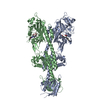
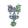
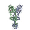
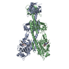
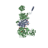
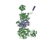
| Title | Crystal structure of the dark-adapted full-length bacteriophytochrome XccBphP from Xanthomonas campestris bound to BV chromophore | ||||||
 Components Components | PHYTOCHROME-LIKE PROTEIN | ||||||
 Keywords Keywords | SIGNALING PROTEIN / PHOTORECEPTOR / BACTERIAL PROTEIN / PHOTOSENSOR / RED/FAR-RED LIGHT / PHYTOCHROME / PROTEIN STRUCTURE / SIGNAL TRANSDUCTION / XANTHOMONAS / PHYTOPATHOGEN | ||||||
| Function / homology |  Function and homology information Function and homology information: / : / detection of visible light / photoreceptor activity / regulation of DNA-templated transcription Similarity search - Function | ||||||
| Biological species |  XANTHOMONAS CAMPESTRIS PV. CAMPESTRIS (bacteria) XANTHOMONAS CAMPESTRIS PV. CAMPESTRIS (bacteria) | ||||||
| Method |  X-RAY DIFFRACTION / SYNCHROTRON / MOLECULAR REPLACEMENT / Resolution: 3.25 Å X-RAY DIFFRACTION / SYNCHROTRON / MOLECULAR REPLACEMENT / Resolution: 3.25 Å | ||||||
 Authors Authors | Otero, L.H. / Klinke, S. / Goldbaum, F.A. / Bonomi, H.R. | ||||||
 Citation Citation | Journal: J.Mol.Biol. / Year: 2016 Title: Structure of the Full-Length Bacteriophytochrome from the Plant Pathogen Xanthomonas Campestris Provides Clues to its Long-Range Signaling Mechanism. Authors: Otero, L.H. / Klinke, S. / Rinaldi, J. / Velazquez-Escobar, F. / Mroginski, M.A. / Lopez, M.F. / Malamud, F. / Vojnov, A.A. / Hildebrandt, P. / Goldbaum, F.A. / Bonomi, H.R. | ||||||
| History |
|
- Structure visualization
| Structure viewer | Molecule: MolmilJmol/JSmol |
|---|
- Downloads & links
-Download
| PDBx/mmCIF format | 5akp.cif.gz | 489.2 KB | Display | PDBx/mmCIF format |
|---|---|---|---|---|
| PDB format | pdb5akp.ent.gz | 410.7 KB | Display | PDB format |
| PDBx/mmJSON format | 5akp.json.gz | Tree view | PDBx/mmJSON format | |
| Others |  Other downloads Other downloads |
-Validation report
| Arichive directory | https://data.pdbj.org/pub/pdb/validation_reports/ak/5akpftp://data.pdbj.org/pub/pdb/validation_reports/ak/5akp | HTTPS FTP |
|---|
-Related structure data
| Related structure data |  4gw9S S: Starting model for refinement |
|---|---|
| Similar structure data |


-Links
 PDBj
PDBj


- Assembly
| Deposited unit | 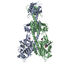
| ||||||||
|---|---|---|---|---|---|---|---|---|---|
| 1 |
| ||||||||
| Unit cell |
|
-Components
| #1: Protein | Mass: 71266.172 Da / Num. of mol.: 2 Source method: isolated from a genetically manipulated source Details: THIOETHER BOND BETWEEN THE A-RING CBC SIDE CHAIN FROM BV AND THE CYS 13 Source: (gene. exp.) XANTHOMONAS CAMPESTRIS PV. CAMPESTRIS (bacteria)Strain: 8004 / Plasmid: PET-24A / Production host: #2: Chemical |   Mass: 582.646 Da / Num. of mol.: 2 / Source method: obtained synthetically / Formula: C33H34N4O6 Mass: 582.646 Da / Num. of mol.: 2 / Source method: obtained synthetically / Formula: C33H34N4O6#3: Chemical |   Mass: 122.143 Da / Num. of mol.: 2 / Source method: obtained synthetically / Formula: C4H12NO3 / Comment: pH buffer*YM Mass: 122.143 Da / Num. of mol.: 2 / Source method: obtained synthetically / Formula: C4H12NO3 / Comment: pH buffer*YM#4: Chemical | ChemComp-CL / |   Mass: 35.453 Da / Num. of mol.: 1 / Source method: obtained synthetically / Formula: Cl Mass: 35.453 Da / Num. of mol.: 1 / Source method: obtained synthetically / Formula: Cl#5: Water | ChemComp-HOH / |  Mass: 18.015 Da / Num. of mol.: 2 / Source method: isolated from a natural source / Formula: H2O Mass: 18.015 Da / Num. of mol.: 2 / Source method: isolated from a natural source / Formula: H2OHas protein modification | Y | |
|---|
-Experimental details
-Experiment
| Experiment | Method: X-RAY DIFFRACTION / Number of used crystals: 1 |
|---|
- Sample preparation
| Crystal | Density Matthews: 3.27 Å3/Da / Density % sol: 62 % / Description: NONE |
|---|---|
| Crystal grow | pH: 8.3 Details: 12%(W/V) PEG 4000, 0.1 M TRIS, 0.2 M SODIUM ACETATE PH 8.3 |
-Data collection
| Diffraction | Mean temperature: 100 K |
|---|---|
| Diffraction source | Source: SYNCHROTRON / Site: SOLEIL  / Beamline: PROXIMA 1 / Wavelength: 0.97857 / Beamline: PROXIMA 1 / Wavelength: 0.97857 |
| Detector | Type: DECTRIS PILATUS 6M / Detector: PIXEL / Date: Nov 24, 2013 / Details: KIRKPATRICK-BAEZ PAIR OF BI-MORPH MIRRORS |
| Radiation | Monochromator: CHANNEL CUT CRYOGENICALLY COOLED MONOCROMATOR CRYSTAL Protocol: SINGLE WAVELENGTH / Monochromatic (M) / Laue (L): M / Scattering type: x-ray |
| Radiation wavelength | Wavelength: 0.97857 Å / Relative weight: 1 |
| Reflection | Resolution: 3.25→47.35 Å / Num. obs: 30865 / % possible obs: 99.9 % / Observed criterion σ(I): 2 / Redundancy: 7 % / Biso Wilson estimate: 94.46 Å2 / Rmerge(I) obs: 0.12 / Net I/σ(I): 19.6 |
| Reflection shell | Resolution: 3.25→3.43 Å / Redundancy: 7.5 % / Rmerge(I) obs: 1.49 / Mean I/σ(I) obs: 2.4 / % possible all: 100 |
- Processing
| Software |
| ||||||||||||||||||||||||||||||||||||||||||||||||||||||||||||||||||||||||||||||||||||||||||||||||||||||||||||||||||
|---|---|---|---|---|---|---|---|---|---|---|---|---|---|---|---|---|---|---|---|---|---|---|---|---|---|---|---|---|---|---|---|---|---|---|---|---|---|---|---|---|---|---|---|---|---|---|---|---|---|---|---|---|---|---|---|---|---|---|---|---|---|---|---|---|---|---|---|---|---|---|---|---|---|---|---|---|---|---|---|---|---|---|---|---|---|---|---|---|---|---|---|---|---|---|---|---|---|---|---|---|---|---|---|---|---|---|---|---|---|---|---|---|---|---|---|
| Refinement | Method to determine structure: MOLECULAR REPLACEMENT Starting model: PDB ENTRY 4GW9 Resolution: 3.25→41.75 Å / Cor.coef. Fo:Fc: 0.9433 / Cor.coef. Fo:Fc free: 0.9049 / Cross valid method: THROUGHOUT / σ(F): 0 / SU Rfree Blow DPI: 0.453 Details: 11 N-TERMINAL RESIDUES AND RESIDUES FROM 456 TO 470 NOT MODELED, WEAK ELECTRON DENSITY IS SHOWN AT THE AREA WHERE THE D-RING FROM BV CHROMOPHORE IS PLACED.
| ||||||||||||||||||||||||||||||||||||||||||||||||||||||||||||||||||||||||||||||||||||||||||||||||||||||||||||||||||
| Displacement parameters | Biso mean: 114.59 Å2
| ||||||||||||||||||||||||||||||||||||||||||||||||||||||||||||||||||||||||||||||||||||||||||||||||||||||||||||||||||
| Refine analyze | Luzzati coordinate error obs: 0.846 Å | ||||||||||||||||||||||||||||||||||||||||||||||||||||||||||||||||||||||||||||||||||||||||||||||||||||||||||||||||||
| Refinement step | Cycle: LAST / Resolution: 3.25→41.75 Å
| ||||||||||||||||||||||||||||||||||||||||||||||||||||||||||||||||||||||||||||||||||||||||||||||||||||||||||||||||||
| Refine LS restraints |
| ||||||||||||||||||||||||||||||||||||||||||||||||||||||||||||||||||||||||||||||||||||||||||||||||||||||||||||||||||
| LS refinement shell | Resolution: 3.25→3.36 Å / Total num. of bins used: 15
| ||||||||||||||||||||||||||||||||||||||||||||||||||||||||||||||||||||||||||||||||||||||||||||||||||||||||||||||||||
| Refinement TLS params. | Method: refined / Refine-ID: X-RAY DIFFRACTION
|
