+ Open data
- Basic information
| Entry | Database: PDB / ID: 5aj2 | ||||||
|---|---|---|---|---|---|---|---|
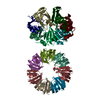
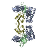
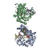
| Title | Cryo electron tomography of the Naip5-Nlrc4 inflammasome | ||||||
 Components Components | (NLR FAMILY CARD DOMAIN-CONTAINING PROTEIN 4) x 3 | ||||||
 Keywords Keywords | APOPTOSIS | ||||||
| Function / homology |  Function and homology information Function and homology informationIPAF inflammasome complex / caspase binding / positive regulation of protein processing / pyroptotic inflammatory response / endopeptidase activator activity / detection of bacterium / activation of innate immune response / positive regulation of interleukin-1 beta production / protein homooligomerization / regulation of apoptotic process ...IPAF inflammasome complex / caspase binding / positive regulation of protein processing / pyroptotic inflammatory response / endopeptidase activator activity / detection of bacterium / activation of innate immune response / positive regulation of interleukin-1 beta production / protein homooligomerization / regulation of apoptotic process / defense response to bacterium / positive regulation of apoptotic process / inflammatory response / innate immune response / intracellular membrane-bounded organelle / apoptotic process / protein homodimerization activity / ATP binding / identical protein binding / plasma membrane / cytosol Similarity search - Function | ||||||
| Biological species |  | ||||||
| Method | ELECTRON MICROSCOPY / electron tomography / cryo EM / Resolution: 40 Å | ||||||
| Model type details | CA ATOMS ONLY, CHAIN A, C, B | ||||||
 Authors Authors | Diebolder, C.A. / Halff, E.F. / Koster, A.J. / Huizinga, E.G. / Koning, R.I. | ||||||
 Citation Citation |  Journal: Structure / Year: 2015 Journal: Structure / Year: 2015Title: Cryoelectron Tomography of the NAIP5/NLRC4 Inflammasome: Implications for NLR Activation. Authors: Christoph A Diebolder / Els F Halff / Abraham J Koster / Eric G Huizinga / Roman I Koning /  Abstract: Inflammasomes are high molecular weight protein complexes that play a crucial role in innate immunity by activating caspase-1. Inflammasome formation is initiated when molecules originating from ...Inflammasomes are high molecular weight protein complexes that play a crucial role in innate immunity by activating caspase-1. Inflammasome formation is initiated when molecules originating from invading microorganisms activate nucleotide-binding domain and leucine-rich repeat-containing receptors (NLRs) and induce NLR multimerization. Little is known about the conformational changes involved in NLR activation and the structural organization of NLR multimers. Here, we show by cryoelectron tomography that flagellin-induced NAIP5/NLRC4 multimers form right- and left-handed helical polymers with a diameter of 28 nm and a pitch of 6.5 nm. Subtomogram averaging produced an electron density map at 4 nm resolution, which was used for rigid body fitting of NLR subdomains derived from the crystal structure of dormant NLRC4. The resulting structural model of inflammasome-incorporated NLRC4 indicates that a prominent rotation of the LRR domain of NLRC4 is necessary for multimer formation, providing unprecedented insight into the conformational changes that accompany NLR activation. | ||||||
| History |
|
- Structure visualization
| Movie |
 Movie viewer Movie viewer |
|---|---|
| Structure viewer | Molecule:  Molmil Molmil Jmol/JSmol Jmol/JSmol |
- Downloads & links
- Download
| PDBx/mmCIF format |  5aj2.cif.gz 5aj2.cif.gz | 48.8 KB | Display |  PDBx/mmCIF format PDBx/mmCIF format |
|---|---|---|---|---|
| PDB format |  pdb5aj2.ent.gz pdb5aj2.ent.gz | 24.5 KB | Display |  PDB format PDB format |
| PDBx/mmJSON format |  5aj2.json.gz 5aj2.json.gz | Tree view |  PDBx/mmJSON format PDBx/mmJSON format | |
| Others |  Other downloads Other downloads |
-Validation report
| Summary document |  5aj2_validation.pdf.gz 5aj2_validation.pdf.gz | 522.5 KB | Display |  wwPDB validaton report wwPDB validaton report |
|---|---|---|---|---|
| Full document |  5aj2_full_validation.pdf.gz 5aj2_full_validation.pdf.gz | 524.2 KB | Display | |
| Data in XML |  5aj2_validation.xml.gz 5aj2_validation.xml.gz | 16.2 KB | Display | |
| Data in CIF |  5aj2_validation.cif.gz 5aj2_validation.cif.gz | 23.9 KB | Display | |
| Arichive directory |  https://data.pdbj.org/pub/pdb/validation_reports/aj/5aj2 https://data.pdbj.org/pub/pdb/validation_reports/aj/5aj2 ftp://data.pdbj.org/pub/pdb/validation_reports/aj/5aj2 ftp://data.pdbj.org/pub/pdb/validation_reports/aj/5aj2 | HTTPS FTP |
-Related structure data
| Related structure data |  2901MC M: map data used to model this data C: citing same article ( |
|---|---|
| Similar structure data |
- Links
Links
- Assembly
| Deposited unit | 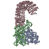
|
|---|---|
| 1 |
|
| Noncrystallographic symmetry (NCS) | NCS oper: (Code: given / Matrix: (0.858065, 0.513541), |
- Components
| #1: Protein | Mass: 40914.316 Da / Num. of mol.: 1 / Fragment: RESIDUES 1-355 Source method: isolated from a genetically manipulated source Source: (gene. exp.)   HOMO SAPIENS (human) / References: UniProt: Q3UP24 HOMO SAPIENS (human) / References: UniProt: Q3UP24 |
|---|---|
| #2: Protein | Mass: 25524.809 Da / Num. of mol.: 1 / Fragment: RESIDUES 356-580 Source method: isolated from a genetically manipulated source Source: (gene. exp.)   HOMO SAPIENS (human) / References: UniProt: Q3UP24 HOMO SAPIENS (human) / References: UniProt: Q3UP24 |
| #3: Protein | Mass: 50610.996 Da / Num. of mol.: 1 / Fragment: RESIDUES 580-1024 Source method: isolated from a genetically manipulated source Source: (gene. exp.)   HOMO SAPIENS (human) / References: UniProt: Q3UP24 HOMO SAPIENS (human) / References: UniProt: Q3UP24 |
-Experimental details
-Experiment
| Experiment | Method: ELECTRON MICROSCOPY |
|---|---|
| EM experiment | Aggregation state: HELICAL ARRAY / 3D reconstruction method: electron tomography |
- Sample preparation
| Component | Name: NAIP5-NLRC4-FLIC-D0L MULTIMER / Type: COMPLEX |
|---|---|
| Buffer solution | Name: 100 MM NACL, 20 MM HEPES, 2MM BENZAMIDIN, 2MM DTT / pH: 7.5 / Details: 100 MM NACL, 20 MM HEPES, 2MM BENZAMIDIN, 2MM DTT |
| Specimen | Conc.: 1.2 mg/ml / Embedding applied: NO / Shadowing applied: NO / Staining applied: NO / Vitrification applied: YES |
| Specimen support | Details: HOLEY CARBON |
| Vitrification | Instrument: LEICA EM GP / Cryogen name: ETHANE-PROPANE Details: VITRIFICATION 1 -- CRYOGEN- ETHANE-PROPANE MIXTURE, HUMIDITY- 95, INSTRUMENT- LEICA EM GP, METHOD- 3 SECONDS BLOTTING, |
- Electron microscopy imaging
| Experimental equipment |  Model: Titan Krios / Image courtesy: FEI Company |
|---|---|
| Microscopy | Model: FEI TITAN KRIOS / Date: May 21, 2013 |
| Electron gun | Electron source:  FIELD EMISSION GUN / Accelerating voltage: 200 kV / Illumination mode: FLOOD BEAM FIELD EMISSION GUN / Accelerating voltage: 200 kV / Illumination mode: FLOOD BEAM |
| Electron lens | Mode: BRIGHT FIELD / Nominal magnification: 18000 X / Nominal defocus max: 7500 nm / Nominal defocus min: 6500 nm |
| Specimen holder | Tilt angle max: 66 ° / Tilt angle min: -66 ° |
| Image recording | Electron dose: 100 e/Å2 / Film or detector model: GATAN ULTRASCAN 4000 (4k x 4k) |
| Image scans | Num. digital images: 67 |
- Processing
| EM software |
| ||||||||||||
|---|---|---|---|---|---|---|---|---|---|---|---|---|---|
| CTF correction | Details: TOMOCTF | ||||||||||||
| 3D reconstruction | Method: CROSS-COMMON LINES / Resolution: 40 Å / Num. of particles: 50 / Nominal pixel size: 5.463 Å / Actual pixel size: 5.463 Å Details: SUBMISSION BASED ON EXPERIMENTAL DATA FROM EMDB EMD-2901. (DEPOSITION ID: 13118). Symmetry type: HELICAL | ||||||||||||
| Atomic model building | Protocol: RIGID BODY FIT / Space: REAL / Details: METHOD--RIGID BODY REFINEMENT PROTOCOL--X-RAY | ||||||||||||
| Atomic model building | PDB-ID: 4KXF Accession code: 4KXF / Source name: PDB / Type: experimental model | ||||||||||||
| Refinement | Highest resolution: 40 Å | ||||||||||||
| Refinement step | Cycle: LAST / Highest resolution: 40 Å
|
 Movie
Movie Controller




 PDBj
PDBj


