+ データを開く
- 基本情報
基本情報
| 登録情報 | データベース: PDB / ID: 5a9e | ||||||
|---|---|---|---|---|---|---|---|
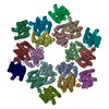
| タイトル | Cryo-electron tomography and subtomogram averaging of Rous-Sarcoma- Virus deltaMBD virus-like particles | ||||||
 要素 要素 | DELTAMBD GAG PROTEIN | ||||||
 キーワード キーワード | VIRAL PROTEIN / RETROVIRUS / ROUS-SARCOMA VIRUS / IMMATURE RETROVIRUS / VIRUS-LIKE-PARTICLE / CAPSID | ||||||
| 機能・相同性 |  機能・相同性情報 機能・相同性情報host cell nucleoplasm / viral procapsid maturation / host cell nucleolus / 加水分解酵素; プロテアーゼ; ペプチド結合加水分解酵素; アスパラギン酸プロテアーゼ / viral capsid / structural constituent of virion / nucleic acid binding / aspartic-type endopeptidase activity / viral translational frameshifting / host cell plasma membrane ...host cell nucleoplasm / viral procapsid maturation / host cell nucleolus / 加水分解酵素; プロテアーゼ; ペプチド結合加水分解酵素; アスパラギン酸プロテアーゼ / viral capsid / structural constituent of virion / nucleic acid binding / aspartic-type endopeptidase activity / viral translational frameshifting / host cell plasma membrane / proteolysis / zinc ion binding / membrane 類似検索 - 分子機能 | ||||||
| 生物種 |  ROUS SARCOMA VIRUS (ラウス肉腫ウイルス) ROUS SARCOMA VIRUS (ラウス肉腫ウイルス) | ||||||
| 手法 | 電子顕微鏡法 / 電子線トモグラフィー法 / クライオ電子顕微鏡法 / 解像度: 7.7 Å | ||||||
 データ登録者 データ登録者 | Schur, F.K.M. / Dick, R.A. / Hagen, W.J.H. / Vogt, V.M. / Briggs, J.A.G. | ||||||
 引用 引用 |  ジャーナル: J Virol / 年: 2015 ジャーナル: J Virol / 年: 2015タイトル: The Structure of Immature Virus-Like Rous Sarcoma Virus Gag Particles Reveals a Structural Role for the p10 Domain in Assembly. 著者: Florian K M Schur / Robert A Dick / Wim J H Hagen / Volker M Vogt / John A G Briggs /   要旨: The polyprotein Gag is the primary structural component of retroviruses. Gag consists of independently folded domains connected by flexible linkers. Interactions between the conserved capsid (CA) ...The polyprotein Gag is the primary structural component of retroviruses. Gag consists of independently folded domains connected by flexible linkers. Interactions between the conserved capsid (CA) domains of Gag mediate formation of hexameric protein lattices that drive assembly of immature virus particles. Proteolytic cleavage of Gag by the viral protease (PR) is required for maturation of retroviruses from an immature form into an infectious form. Within the assembled Gag lattices of HIV-1 and Mason-Pfizer monkey virus (M-PMV), the C-terminal domain of CA adopts similar quaternary arrangements, while the N-terminal domain of CA is packed in very different manners. Here, we have used cryo-electron tomography and subtomogram averaging to study in vitro-assembled, immature virus-like Rous sarcoma virus (RSV) Gag particles and have determined the structure of CA and the surrounding regions to a resolution of ∼8 Å. We found that the C-terminal domain of RSV CA is arranged similarly to HIV-1 and M-PMV, whereas the N-terminal domain of CA adopts a novel arrangement in which the upstream p10 domain folds back into the CA lattice. In this position the cleavage site between CA and p10 appears to be inaccessible to PR. Below CA, an extended density is consistent with the presence of a six-helix bundle formed by the spacer-peptide region. We have also assessed the affect of lattice assembly on proteolytic processing by exogenous PR. The cleavage between p10 and CA is indeed inhibited in the assembled lattice, a finding consistent with structural regulation of proteolytic maturation. IMPORTANCE: Retroviruses first assemble into immature virus particles, requiring interactions between Gag proteins that form a protein layer under the viral membrane. Subsequently, Gag is cleaved by ...IMPORTANCE: Retroviruses first assemble into immature virus particles, requiring interactions between Gag proteins that form a protein layer under the viral membrane. Subsequently, Gag is cleaved by the viral protease enzyme into separate domains, leading to rearrangement of the virus into its infectious form. It is important to understand how Gag is arranged within immature retroviruses, in order to understand how virus assembly occurs, and how maturation takes place. We used the techniques cryo-electron tomography and subtomogram averaging to obtain a detailed structural picture of the CA domains in immature assembled Rous sarcoma virus Gag particles. We found that part of Gag next to CA, called p10, folds back and interacts with CA when Gag assembles. This arrangement is different from that seen in HIV-1 and Mason-Pfizer monkey virus, illustrating further structural diversity of retroviral structures. The structure provides new information on how the virus assembles and undergoes maturation. | ||||||
| 履歴 |
|
- 構造の表示
構造の表示
| ムービー |
 ムービービューア ムービービューア |
|---|---|
| 構造ビューア | 分子:  Molmil Molmil Jmol/JSmol Jmol/JSmol |
- ダウンロードとリンク
- ダウンロード
| PDBx/mmCIF形式 |  5a9e.cif.gz 5a9e.cif.gz | 561.8 KB | 表示 |  PDBx/mmCIF形式 PDBx/mmCIF形式 |
|---|---|---|---|---|
| PDB形式 |  pdb5a9e.ent.gz pdb5a9e.ent.gz | 328.3 KB | 表示 |  PDB形式 PDB形式 |
| PDBx/mmJSON形式 |  5a9e.json.gz 5a9e.json.gz | ツリー表示 |  PDBx/mmJSON形式 PDBx/mmJSON形式 | |
| その他 |  その他のダウンロード その他のダウンロード |
-検証レポート
| 文書・要旨 |  5a9e_validation.pdf.gz 5a9e_validation.pdf.gz | 769.9 KB | 表示 |  wwPDB検証レポート wwPDB検証レポート |
|---|---|---|---|---|
| 文書・詳細版 |  5a9e_full_validation.pdf.gz 5a9e_full_validation.pdf.gz | 806.8 KB | 表示 | |
| XML形式データ |  5a9e_validation.xml.gz 5a9e_validation.xml.gz | 87 KB | 表示 | |
| CIF形式データ |  5a9e_validation.cif.gz 5a9e_validation.cif.gz | 148.9 KB | 表示 | |
| アーカイブディレクトリ |  https://data.pdbj.org/pub/pdb/validation_reports/a9/5a9e https://data.pdbj.org/pub/pdb/validation_reports/a9/5a9e ftp://data.pdbj.org/pub/pdb/validation_reports/a9/5a9e ftp://data.pdbj.org/pub/pdb/validation_reports/a9/5a9e | HTTPS FTP |
-関連構造データ
- リンク
リンク
- 集合体
集合体
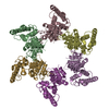
| 登録構造単位 | 
|
|---|---|
| 1 | 
|
| 2 | 
|
| 3 | 
|
| 4 | 
|
| 5 | 
|
| 6 | 
|
| 7 | 
|
- 要素
要素
| #1: タンパク質 | 分子量: 52373.262 Da / 分子数: 18 / 由来タイプ: 組換発現 由来: (組換発現)  ROUS SARCOMA VIRUS (ラウス肉腫ウイルス) ROUS SARCOMA VIRUS (ラウス肉腫ウイルス)発現宿主:  |
|---|
-実験情報
-実験
| 実験 | 手法: 電子顕微鏡法 |
|---|---|
| EM実験 | 試料の集合状態: PARTICLE / 3次元再構成法: 電子線トモグラフィー法 |
- 試料調製
試料調製
| 構成要素 | 名称: IMMATURE ROUS-SARCOMA VIRUS GAG PARTICLES / タイプ: VIRUS |
|---|---|
| 緩衝液 | 名称: MES PH6.5, 100MM NACL, 2UM ZNCL2, 2MM TCEP / pH: 6.5 / 詳細: MES PH6.5, 100MM NACL, 2UM ZNCL2, 2MM TCEP |
| 試料 | 濃度: 5 mg/ml / 包埋: NO / シャドウイング: NO / 染色: NO / 凍結: YES |
| 試料支持 | 詳細: HOLEY CARBON |
| 急速凍結 | 装置: FEI VITROBOT MARK II / 凍結剤: ETHANE 詳細: DEGASSED C-FLAT 2 2-3C GRIDS WERE GLOW DISCHARGED FOR 30 SECONDS AT 20 MA. VIRUS SOLUTION WAS DILUTED IN OBS CONTAINING 10NM COLLOIDAL GOLD. 2.5UL OF THIS MIXTURE WAS APPLIED TO A GRID. BLOTTING TIME 2.5 SECONDS. |
- 電子顕微鏡撮影
| 実験機器 |  モデル: Titan Krios / 画像提供: FEI Company |
|---|---|
| 顕微鏡 | モデル: FEI TITAN KRIOS / 日付: 2014年9月10日 |
| 電子銃 | 電子線源:  FIELD EMISSION GUN / 加速電圧: 200 kV / 照射モード: FLOOD BEAM FIELD EMISSION GUN / 加速電圧: 200 kV / 照射モード: FLOOD BEAM |
| 電子レンズ | モード: BRIGHT FIELD / 倍率(公称値): 81000 X / 最大 デフォーカス(公称値): 5000 nm / 最小 デフォーカス(公称値): 1500 nm / Cs: 2.7 mm |
| 試料ホルダ | 傾斜角・最大: 45 ° / 傾斜角・最小: -45 ° |
| 撮影 | 電子線照射量: 34 e/Å2 / フィルム・検出器のモデル: GATAN MULTISCAN |
| 画像スキャン | デジタル画像の数: 31 |
| 放射波長 | 相対比: 1 |
- 解析
解析
| EMソフトウェア |
| ||||||||||||||||||||||||
|---|---|---|---|---|---|---|---|---|---|---|---|---|---|---|---|---|---|---|---|---|---|---|---|---|---|
| CTF補正 | 詳細: PHASE-FLIPPING OF INDIVIDUAL MICROGRAPHS | ||||||||||||||||||||||||
| 対称性 | 点対称性: C6 (6回回転対称) | ||||||||||||||||||||||||
| 3次元再構成 | 手法: CROSS-CORRELATION / 解像度: 7.7 Å / 粒子像の数: 8375 / ピクセルサイズ(公称値): 2.07 Å / ピクセルサイズ(実測値): 2.07 Å 詳細: RECONSTRUCTION CARRIED OUT USING SUBTOMOGRAM AVERAGING. SUBTOMOGRAM AVERAGING WAS PERFORMED USING SCRIPTS DERIVED FROM THE AV3 (FOERSTER, ET AL) TOM (NICKELL, ET AL) AND DYNAMO (CASTANO-DIEZ, ...詳細: RECONSTRUCTION CARRIED OUT USING SUBTOMOGRAM AVERAGING. SUBTOMOGRAM AVERAGING WAS PERFORMED USING SCRIPTS DERIVED FROM THE AV3 (FOERSTER, ET AL) TOM (NICKELL, ET AL) AND DYNAMO (CASTANO-DIEZ, ET AL) PACKAGES. STRUCTURES FOR P10 AND CA-NTD (PDB 1P7N) AND CA-CTD (PDB 3G1I, ONE MONOMER) WERE RIGID BODY DOCKED INTO THE EM-MAP USING THE FIT-IN-MAP OPTION IN CHIMERA. REDUNDANT RESIDUES OF THE ANTIPARALLEL DIMER OF PDB 1P7N WERE REMOVED. MISSING RESIDUES IN THE HELIX 7-HELIX 8 LINKER REGION OF CA, THE CONNECTION LOOP BETWEEN P10 AND CA-NTD AND RESIDUES UPSTREAM OF THE HELIX IN P10 WERE MANUALLY MODELED USING COOT. THE FIT WAS FURTHER REFINED USING MOLECULAR DYNAMICS FLEXIBLE FITTING. SUBMISSION BASED ON EXPERIMENTAL DATA FROM EMDB EMD-3101. (DEPOSITION ID: 13603). 対称性のタイプ: POINT | ||||||||||||||||||||||||
| 原子モデル構築 | プロトコル: FLEXIBLE FIT / 空間: REAL / 詳細: METHOD--MOLECULAR DYNAMICS FLEXIBLE FITTING | ||||||||||||||||||||||||
| 精密化 | 最高解像度: 7.7 Å | ||||||||||||||||||||||||
| 精密化ステップ | サイクル: LAST / 最高解像度: 7.7 Å
|
 ムービー
ムービー コントローラー


 PDBj
PDBj