Mass: 35933.828 Da / Num. of mol.: 1 / Mutation: YES Source method: isolated from a genetically manipulated source Details: RESIDUES 3-32 AT THE N-TERMINUS AND RESIDUES 244-271 OF THE THIRD INTRACELLULAR LOOP WERE DELETED FROM THE CONSTRUCT. THE CONSTRUCT WAS TRUNCATED AFTER RESIDUE 367 AND A HEXAHIS TAG ADDED. Source: (gene. exp.) MELEAGRIS GALLOPAVO (turkey) / Cell: ERYTHROCYTE / Plasmid: PBACPAK8 / Cell line (production host): High Five / Production host: TRICHOPLUSIA NI (cabbage looper) / References: UniProt: P07700
Mass: 18.015 Da / Num. of mol.: 26 / Source method: isolated from a natural source / Formula: H2O
-
Details
Has protein modification
Y
Sequence details
THE FOLLOWING MUTATIONS WERE MADE TO IMPROVE THERMOSTABILITY ...THE FOLLOWING MUTATIONS WERE MADE TO IMPROVE THERMOSTABILITY R68S,M90V,I129V,Y227A,A282L,F327A,F338M, Y343L. THE FOLLOWING MUTATIONS WERE MADE TO IMPROVE EXPRESSION AND HELP CRYSTALLIZATION C116L, E130W,C358A.
-
Experimental details
-
Experiment
Experiment
Method: X-RAY DIFFRACTION / Number of used crystals: 4
-
Sample preparation
Crystal
Density Matthews: 2.17 Å3/Da / Density % sol: 43.4 % / Description: NONE
Crystal grow
Temperature: 293 K / Method: lipidic cubic phase / pH: 7 Details: 25% PEG600, 0.1M ADA PH7.0, LIPIDIC CUBIC PHASE (LCP), TEMPERATURE 293K
-
Data collection
Diffraction
ID
Mean temperature (K)
Crystal-ID
1
100
1
2
100
1
Diffraction source
Source
Site
Beamline
ID
Wavelength
SYNCHROTRON
ESRF
ID29
1
0.8726
SYNCHROTRON
Diamond
I24
2
0.969
Detector
Type
ID
Detector
PILATUS
1
PIXEL
ADSC CCD
2
CCD
Radiation
ID
Protocol
Monochromatic (M) / Laue (L)
Scattering type
Wavelength-ID
1
SINGLEWAVELENGTH
M
x-ray
1
2
SINGLEWAVELENGTH
M
x-ray
1
Radiation wavelength
ID
Wavelength (Å)
Relative weight
1
0.8726
1
2
0.969
1
Reflection
Resolution: 2.4→37.8 Å / Num. obs: 12526 / % possible obs: 98.4 % / Observed criterion σ(I): 2 / Redundancy: 4.8 % / Rmerge(I) obs: 0.16 / Net I/σ(I): 8.2
Reflection shell
Resolution: 2.4→2.53 Å / Redundancy: 4.9 % / Rmerge(I) obs: 0.7 / Mean I/σ(I) obs: 1.9 / % possible all: 98.3
-
Processing
Software
Name
Version
Classification
REFMAC
5.8.0107
refinement
MOSFLM
datareduction
Aimless
datascaling
PHASER
phasing
Refinement
Method to determine structure: MIR Starting model: PDB ENTRY 4BVN
Resolution: 2.4→37.79 Å / Cor.coef. Fo:Fc: 0.922 / Cor.coef. Fo:Fc free: 0.905 / SU B: 9.816 / SU ML: 0.223 / Cross valid method: THROUGHOUT / ESU R: 0.561 / ESU R Free: 0.275 / Stereochemistry target values: MAXIMUM LIKELIHOOD Details: HYDROGENS HAVE BEEN ADDED IN THE RIDING POSITIONS. U VALUES REFINED INDIVIDUALLY
Rfactor
Num. reflection
% reflection
Selection details
Rfree
0.2479
608
4.8 %
RANDOM
Rwork
0.21683
-
-
-
obs
0.21835
11942
97.99 %
-
Solvent computation
Ion probe radii: 0.8 Å / Shrinkage radii: 0.8 Å / VDW probe radii: 1.2 Å / Solvent model: MASK
 Movie
Movie Controller

 Yorodumi
 Open data
 Basic information
 Components
 Keywords
 Function and homology information
 X-RAY DIFFRACTION /
  Authors
 Citation
 Structure visualization
 Downloads & links
 Other downloads
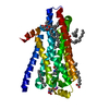


 PDBj
PDBj


 Assembly
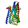
 TRICHOPLUSIA NI (cabbage looper) / References: UniProt: P07700


 Mass: 22.990 Da / Num. of mol.: 2 / Source method: obtained synthetically / Formula: Na
 Mass: 301.383 Da / Num. of mol.: 1 / Source method: obtained synthetically / Formula: C17H23N3O2
 Mass: 190.154 Da / Num. of mol.: 1 / Source method: obtained synthetically / Formula: C6H10N2O5 / Comment: pH buffer*YM
 Mass: 356.540 Da / Num. of mol.: 5 / Source method: obtained synthetically / Formula: C21H40O4
 Sample preparation

 Processing