 Movie
Movie Controller

[English] 日本語
 Yorodumi
- PDB-5a7j: Crystal structure of INPP5B in complex with benzene 1,2,4,5- tetr... -
+ Open data

- Basic information
| Entry | Database: PDB / ID: 5a7j | |||||||||
|---|---|---|---|---|---|---|---|---|---|---|
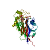
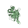
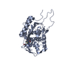
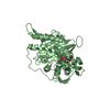
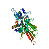
| Title | Crystal structure of INPP5B in complex with benzene 1,2,4,5- tetrakisphosphate | |||||||||
 Components Components | TYPE II INOSITOL 1,4,5-TRISPHOSPHATE 5-PHOSPHATASE | |||||||||
 Keywords Keywords | HYDROLASE / SGC / SIGNALLING / STRUCTURAL GENOMICS CONSORTIUM STOCKHOLM / MAGNESIUM BINDING / PROTEIN-INHBITOR COMPLEX / INHIBITOR / PHOSPHOINOSITIDES SIGNALLING | |||||||||
| Function / homology |  Function and homology information Function and homology informationinositol-1,4,5-trisphosphate 5-phosphatase activity / Synthesis of IP2, IP, and Ins in the cytosol / inositol phosphate metabolic process / inositol-polyphosphate 5-phosphatase / inositol-1,3,4,5-tetrakisphosphate 5-phosphatase activity / phosphoinositide 5-phosphatase / phosphatidylinositol-4,5-bisphosphate 5-phosphatase activity / phosphatidylinositol dephosphorylation / flagellated sperm motility / regulation of protein processing ...inositol-1,4,5-trisphosphate 5-phosphatase activity / Synthesis of IP2, IP, and Ins in the cytosol / inositol phosphate metabolic process / inositol-polyphosphate 5-phosphatase / inositol-1,3,4,5-tetrakisphosphate 5-phosphatase activity / phosphoinositide 5-phosphatase / phosphatidylinositol-4,5-bisphosphate 5-phosphatase activity / phosphatidylinositol dephosphorylation / flagellated sperm motility / regulation of protein processing / endoplasmic reticulum-Golgi intermediate compartment / Synthesis of IP3 and IP4 in the cytosol / ciliary tip / phagocytic vesicle membrane / early endosome membrane / spermatogenesis / in utero embryonic development / neuron projection / Golgi apparatus / signal transduction / membrane / metal ion binding / plasma membrane / cytoplasm / cytosol Similarity search - Function | |||||||||
| Biological species |  HOMO SAPIENS (human) HOMO SAPIENS (human) | |||||||||
| Method |  X-RAY DIFFRACTION / SYNCHROTRON / MOLECULAR REPLACEMENT / Resolution: 2.9 Å X-RAY DIFFRACTION / SYNCHROTRON / MOLECULAR REPLACEMENT / Resolution: 2.9 Å | |||||||||
 Authors Authors | Tresaugues, L. / Mills, S.J. / Silvander, C. / Cozier, G. / Potter, B.V.L. / Nordlund, P. | |||||||||
 Citation Citation | Journal: Biochemistry / Year: 2016 Title: Crystal Structures of Type-II Inositol Polyphosphate 5-Phosphatase Inpp5B with Synthetic Inositol Polyphosphate Surrogates Reveal New Mechanistic Insights for the Inositol 5-Phosphatase Family. Authors: Mills, S.J. / Silvander, C. / Cozier, G. / Tresaugues, L. / Nordlund, P. / Potter, B.V.L. | |||||||||
| History |
|
- Structure visualization
| Structure viewer | Molecule: MolmilJmol/JSmol |
|---|
- Downloads & links
-Download
| PDBx/mmCIF format | 5a7j.cif.gz | 259.3 KB | Display | PDBx/mmCIF format |
|---|---|---|---|---|
| PDB format | pdb5a7j.ent.gz | 213.4 KB | Display | PDB format |
| PDBx/mmJSON format | 5a7j.json.gz | Tree view | PDBx/mmJSON format | |
| Others |  Other downloads Other downloads |
-Validation report
| Arichive directory | https://data.pdbj.org/pub/pdb/validation_reports/a7/5a7jftp://data.pdbj.org/pub/pdb/validation_reports/a7/5a7j | HTTPS FTP |
|---|
-Related structure data
| Related structure data |  5a7iC 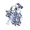 3n9vS C: citing same article ( S: Starting model for refinement |
|---|---|
| Similar structure data |


-Links
 PDBj
PDBj


- Assembly
| Deposited unit | 
| ||||||||
|---|---|---|---|---|---|---|---|---|---|
| 1 | 
| ||||||||
| 2 | 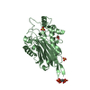
| ||||||||
| Unit cell |
| ||||||||
| Noncrystallographic symmetry (NCS) | NCS oper: (Code: given Matrix: (0.72408, 0.00399, -0.68971), Vector: |
-Components
| #1: Protein | Mass: 36201.086 Da / Num. of mol.: 2 / Fragment: UNP RESIDUES 339-643 Source method: isolated from a genetically manipulated source Details: CLONED WITH A C-TERMINAL HEXAHISTIDINE TAG / Source: (gene. exp.) HOMO SAPIENS (human) / Description: MAMMALIAN GENE COLLECTION (MGC) / Plasmid: PNIC-CH2 / Production host:  #2: Chemical | ChemComp-SO4 /   Mass: 96.063 Da / Num. of mol.: 4 / Source method: obtained synthetically / Formula: SO4 Mass: 96.063 Da / Num. of mol.: 4 / Source method: obtained synthetically / Formula: SO4#3: Chemical |   Mass: 462.029 Da / Num. of mol.: 2 / Source method: obtained synthetically / Formula: C6H10O16P4 Mass: 462.029 Da / Num. of mol.: 2 / Source method: obtained synthetically / Formula: C6H10O16P4#4: Chemical | ChemComp-GOL / |   Mass: 92.094 Da / Num. of mol.: 1 / Source method: obtained synthetically / Formula: C3H8O3 Mass: 92.094 Da / Num. of mol.: 1 / Source method: obtained synthetically / Formula: C3H8O3#5: Water | ChemComp-HOH / |  Mass: 18.015 Da / Num. of mol.: 76 / Source method: isolated from a natural source / Formula: H2O Mass: 18.015 Da / Num. of mol.: 76 / Source method: isolated from a natural source / Formula: H2O |
|---|
-Experimental details
-Experiment
| Experiment | Method: X-RAY DIFFRACTION / Number of used crystals: 1 |
|---|
- Sample preparation
| Crystal | Density Matthews: 2.61 Å3/Da / Density % sol: 52.89 % / Description: NONE |
|---|---|
| Crystal grow | pH: 5.5 Details: 0.2 M LI2SO4, 0.1 M BIS-TRIS PH 5.5 AND 25% PEG3350 |
-Data collection
| Diffraction | Mean temperature: 100 K |
|---|---|
| Diffraction source | Source: SYNCHROTRON / Site: BESSY  / Beamline: 14.1 / Wavelength: 0.91841 / Beamline: 14.1 / Wavelength: 0.91841 |
| Detector | Type: MARMOSAIC 225 mm CCD / Detector: CCD / Date: Nov 1, 2011 / Details: MIRRORS |
| Radiation | Monochromator: SI-111 CRYSTAL / Protocol: SINGLE WAVELENGTH / Monochromatic (M) / Laue (L): M / Scattering type: x-ray |
| Radiation wavelength | Wavelength: 0.91841 Å / Relative weight: 1 |
| Reflection | Resolution: 2.9→48.48 Å / Num. obs: 16223 / % possible obs: 99.7 % / Observed criterion σ(I): -3 / Redundancy: 3.6 % / Biso Wilson estimate: 52.88 Å2 / Rmerge(I) obs: 0.16 / Net I/σ(I): 6.9 |
| Reflection shell | Resolution: 2.9→3.06 Å / Redundancy: 3.6 % / Rmerge(I) obs: 0.58 / Mean I/σ(I) obs: 2.3 / % possible all: 99.7 |
- Processing
| Software |
| ||||||||||||||||||||
|---|---|---|---|---|---|---|---|---|---|---|---|---|---|---|---|---|---|---|---|---|---|
| Refinement | Method to determine structure: MOLECULAR REPLACEMENT Starting model: PDB ENTRY 3N9V Resolution: 2.9→48.48 Å / Cor.coef. Fo:Fc: 0.9009 / Cor.coef. Fo:Fc free: 0.8422 / Cross valid method: THROUGHOUT / σ(F): 0 / Details: REMARK 3 NULL
| ||||||||||||||||||||
| Displacement parameters | Biso mean: 39.21 Å2
| ||||||||||||||||||||
| Refinement step | Cycle: LAST / Resolution: 2.9→48.48 Å
| ||||||||||||||||||||
| LS refinement shell | Resolution: 2.9→3.1 Å / Total num. of bins used: 8
|
