 Movie
Movie Controller

[English] 日本語
 Yorodumi
- PDB-4zym: Structural implications of homo-pyrimidine base pairs on the para... -
+ Open data

- Basic information
| Entry | Database: PDB / ID: 4zym | ||||||||||||||||||||||||||||
|---|---|---|---|---|---|---|---|---|---|---|---|---|---|---|---|---|---|---|---|---|---|---|---|---|---|---|---|---|---|
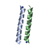
| Title | Structural implications of homo-pyrimidine base pairs on the parallel-stranded d(GAY) motif. | ||||||||||||||||||||||||||||
 Components Components | DNA (5'-D(* Keywords KeywordsDNA / Tetraplex / homo-base pair / pyrimidine substitution. | Function / homology | DNA / DNA (> 10) |  Function and homology information Function and homology informationBiological species | synthetic construct (others) | Method |  X-RAY DIFFRACTION / SYNCHROTRON / MOLECULAR REPLACEMENT / molecular replacement / Resolution: 2.53 Å X-RAY DIFFRACTION / SYNCHROTRON / MOLECULAR REPLACEMENT / molecular replacement / Resolution: 2.53 Å  Authors AuthorsTripathi, S.K. / Paukstelis, P. | Funding support | |  United States, 1items United States, 1items
 CitationJournal: Chembiochem / Year: 2016 CitationJournal: Chembiochem / Year: 2016Title: Structural Implications of Homopyrimidine Base Pairs in the Parallel-Stranded d(YGA) Motif. Authors: Tripathi, S. / Paukstelis, P.J. History |
|
- Structure visualization
| Structure viewer | Molecule: MolmilJmol/JSmol |
|---|
- Downloads & links
-Download
| PDBx/mmCIF format | 4zym.cif.gz | 33 KB | Display | PDBx/mmCIF format |
|---|---|---|---|---|
| PDB format | pdb4zym.ent.gz | 21.7 KB | Display | PDB format |
| PDBx/mmJSON format | 4zym.json.gz | Tree view | PDBx/mmJSON format | |
| Others |  Other downloads Other downloads |
-Validation report
| Summary document | 4zym_validation.pdf.gz | 383.6 KB | Display | wwPDB validaton report |
|---|---|---|---|---|
| Full document | 4zym_full_validation.pdf.gz | 383.6 KB | Display | |
| Data in XML | 4zym_validation.xml.gz | 3.1 KB | Display | |
| Data in CIF | 4zym_validation.cif.gz | 3.8 KB | Display | |
| Arichive directory | https://data.pdbj.org/pub/pdb/validation_reports/zy/4zymftp://data.pdbj.org/pub/pdb/validation_reports/zy/4zym | HTTPS FTP |
-Related structure data
| Related structure data | 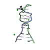 5cv2C  4rimS S: Starting model for refinement C: citing same article ( |
|---|---|
| Similar structure data |

-Links
 PDBj
PDBj


- Assembly
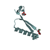
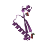
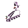
| Deposited unit | 
| ||||||||
|---|---|---|---|---|---|---|---|---|---|
| 1 | 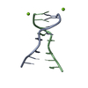
| ||||||||
| 2 | 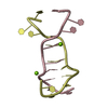
| ||||||||
| Unit cell |
|
-Components
| #1: DNA chain | Mass: 3358.211 Da / Num. of mol.: 4 / Source method: obtained synthetically Details: DNA oligonucleotide demonstrating parallel stranded DNA homoduplex. Source: (synth.) synthetic construct (others) #2: Chemical | ChemComp-MG /   Mass: 24.305 Da / Num. of mol.: 4 / Source method: obtained synthetically / Formula: Mg Mass: 24.305 Da / Num. of mol.: 4 / Source method: obtained synthetically / Formula: Mg#3: Water | ChemComp-HOH / |  Mass: 18.015 Da / Num. of mol.: 6 / Source method: isolated from a natural source / Formula: H2O Mass: 18.015 Da / Num. of mol.: 6 / Source method: isolated from a natural source / Formula: H2O |
|---|
-Experimental details
-Experiment
| Experiment | Method: X-RAY DIFFRACTION / Number of used crystals: 1 |
|---|
- Sample preparation
| Crystal | Density Matthews: 2.26 Å3/Da / Density % sol: 45.62 % / Description: Crystals diffracted to 2.5 Angstrom |
|---|---|
| Crystal grow | Temperature: 300 K / Method: vapor diffusion, sitting drop / pH: 7.5 Details: 100 mM Magnesium chloride, 5 % PEG400, 30 mM Sodium Cacodilate, 8 mM Cobalt hexamine. |
-Data collection
| Diffraction | Mean temperature: 100 K |
|---|---|
| Diffraction source | Source: SYNCHROTRON / Site: APS / Beamline: 24-ID-E / Wavelength: 0.979 Å |
| Detector | Type: ADSC QUANTUM 315 / Detector: CCD / Date: Nov 1, 2014 |
| Radiation | Monochromator: Si(220) / Protocol: SINGLE WAVELENGTH / Monochromatic (M) / Laue (L): M / Scattering type: x-ray |
| Radiation wavelength | Wavelength: 0.979 Å / Relative weight: 1 |
| Reflection | Resolution: 2.5→69.057 Å / Num. all: 11965 / Num. obs: 4342 / % possible obs: 98.73 % / Redundancy: 2.8 % / Biso Wilson estimate: 45.3 Å2 / Rmerge(I) obs: 0.103 / Rsym value: 0.145 / Net I/av σ(I): 6.4 / Net I/σ(I): 4.5 |
| Reflection shell | Resolution: 2.5→2.62 Å / Redundancy: 2.9 % / Rmerge(I) obs: 0.286 / Mean I/σ(I) obs: 1.9 / % possible all: 95 |
-Phasing
| Phasing | Method: molecular replacement | |||||||||
|---|---|---|---|---|---|---|---|---|---|---|
| Phasing MR | Model details: Phaser MODE: MR_AUTO
|
- Processing
| Software |
| ||||||||||||||||||||||||
|---|---|---|---|---|---|---|---|---|---|---|---|---|---|---|---|---|---|---|---|---|---|---|---|---|---|
| Refinement | Method to determine structure: MOLECULAR REPLACEMENT Starting model: 4RIM Resolution: 2.53→69.057 Å / SU ML: 0.26 / Cross valid method: FREE R-VALUE / σ(F): 1.38 / Phase error: 30.69 / Stereochemistry target values: ML
| ||||||||||||||||||||||||
| Solvent computation | Shrinkage radii: 0.9 Å / VDW probe radii: 1.11 Å / Solvent model: FLAT BULK SOLVENT MODEL | ||||||||||||||||||||||||
| Displacement parameters | Biso max: 100.03 Å2 / Biso mean: 35.2971 Å2 / Biso min: 13.71 Å2 | ||||||||||||||||||||||||
| Refinement step | Cycle: final / Resolution: 2.53→69.057 Å
| ||||||||||||||||||||||||
| Refine LS restraints |
| ||||||||||||||||||||||||
| LS refinement shell | Resolution: 2.53→2.768 Å
|
