 Movie
Movie Controller

[English] 日本語
 Yorodumi
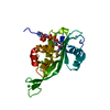
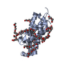
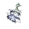
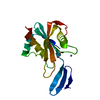
- PDB-4zv5: Crystal structure of N-myristoylated mouse mammary tumor virus ma... -
+ Open data

- Basic information
| Entry | Database: PDB / ID: 4zv5 | ||||||
|---|---|---|---|---|---|---|---|
| Title | Crystal structure of N-myristoylated mouse mammary tumor virus matrix protein | ||||||
 Components Components | Matrix protein p10 | ||||||
 Keywords Keywords | VIRAL PROTEIN / retroviral matrix protein / N-myristoylation | ||||||
| Function / homology |  Function and homology information Function and homology informationviral budding via host ESCRT complex / viral nucleocapsid / structural constituent of virion / viral translational frameshifting / nucleotide binding / DNA binding / zinc ion binding Similarity search - Function | ||||||
| Biological species |  Mouse mammary tumor virus Mouse mammary tumor virus | ||||||
| Method |  X-RAY DIFFRACTION / SYNCHROTRON / SIRAS / Resolution: 1.57 Å X-RAY DIFFRACTION / SYNCHROTRON / SIRAS / Resolution: 1.57 Å | ||||||
 Authors Authors | Zabransky, A. / Dolezal, M. / Dostal, J. / Vanek, O. / Hadravova, R. / Stokrova, J. / Brynda, J. / Pichova, I. | ||||||
 Citation Citation | Journal: Retrovirology / Year: 2016 Title: Myristoylation drives dimerization of matrix protein from mouse mammary tumor virus. Authors: Dolezal, M. / Zabransky, A. / Dostal, J. / Vanek, O. / Brynda, J. / Lepsik, M. / Hadravova, R. / Pichova, I. #1: Journal: Protein Expr.Purif. / Year: 2013 Title: One-step separation of myristoylated and nonmyristoylated retroviral matrix proteins. Authors: Dolezal, M. / Zabransky, A. / Hrabal, R. / Ruml, T. / Pichova, I. / Rumlova, M. | ||||||
| History |
|
- Structure visualization
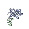
| Structure viewer | Molecule: MolmilJmol/JSmol |
|---|
- Downloads & links
-Download
| PDBx/mmCIF format | 4zv5.cif.gz | 53.1 KB | Display | PDBx/mmCIF format |
|---|---|---|---|---|
| PDB format | pdb4zv5.ent.gz | 38.1 KB | Display | PDB format |
| PDBx/mmJSON format | 4zv5.json.gz | Tree view | PDBx/mmJSON format | |
| Others |  Other downloads Other downloads |
-Validation report
| Arichive directory | https://data.pdbj.org/pub/pdb/validation_reports/zv/4zv5ftp://data.pdbj.org/pub/pdb/validation_reports/zv/4zv5 | HTTPS FTP |
|---|
-Related structure data
| Similar structure data |
|---|


-Links
 PDBj
PDBj


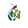
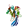
- Assembly
| Deposited unit | 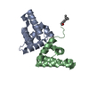
| ||||||||
|---|---|---|---|---|---|---|---|---|---|
| 1 |
| ||||||||
| Unit cell |
|
-Components
| #1: Protein | Mass: 10489.055 Da / Num. of mol.: 2 / Fragment: UNP residues 2-92 Source method: isolated from a genetically manipulated source Source: (gene. exp.) Mouse mammary tumor virus / Gene: gag / Plasmid: pET-22b / Production host:  #2: Chemical |   Mass: 228.371 Da / Num. of mol.: 2 / Source method: obtained synthetically / Formula: C14H28O2 Mass: 228.371 Da / Num. of mol.: 2 / Source method: obtained synthetically / Formula: C14H28O2#3: Water | ChemComp-HOH / |  Mass: 18.015 Da / Num. of mol.: 87 / Source method: isolated from a natural source / Formula: H2O Mass: 18.015 Da / Num. of mol.: 87 / Source method: isolated from a natural source / Formula: H2OHas protein modification | Y | |
|---|
-Experimental details
-Experiment
| Experiment | Method: X-RAY DIFFRACTION / Number of used crystals: 1 |
|---|
- Sample preparation
| Crystal | Density Matthews: 2.38 Å3/Da / Density % sol: 48.24 % |
|---|---|
| Crystal grow | Temperature: 294 K / Method: vapor diffusion, hanging drop / pH: 8 Details: 0.2M potassium chloride, 20% PEG 3350, pH 8.0, VAPOR DIFFUSION, HANGING DROP, temperature 294K |
-Data collection
| Diffraction | Mean temperature: 100 K | ||||||||||||||||||||||||||||||||||||
|---|---|---|---|---|---|---|---|---|---|---|---|---|---|---|---|---|---|---|---|---|---|---|---|---|---|---|---|---|---|---|---|---|---|---|---|---|---|
| Diffraction source | Source: SYNCHROTRON / Site: BESSY  / Beamline: 14.2 / Wavelength: 0.9184 Å / Beamline: 14.2 / Wavelength: 0.9184 Å | ||||||||||||||||||||||||||||||||||||
| Detector | Type: MARMOSAIC 225 mm CCD / Detector: CCD / Date: May 29, 2013 | ||||||||||||||||||||||||||||||||||||
| Radiation | Protocol: SINGLE WAVELENGTH / Monochromatic (M) / Laue (L): M / Scattering type: x-ray | ||||||||||||||||||||||||||||||||||||
| Radiation wavelength | Wavelength: 0.9184 Å / Relative weight: 1 | ||||||||||||||||||||||||||||||||||||
| Reflection | Rmerge(I) obs: 0.036 / D res high: 1.9 Å | ||||||||||||||||||||||||||||||||||||
| Diffraction reflection shell |
| ||||||||||||||||||||||||||||||||||||
| Reflection | Resolution: 1.57→46.08 Å / Num. all: 28486 / Num. obs: 28486 / % possible obs: 99.7 % / Observed criterion σ(F): 0 / Observed criterion σ(I): 0 / Redundancy: 6.2 % / Biso Wilson estimate: 30.3 Å2 / Rmerge(I) obs: 0.039 / Net I/σ(I): 23.9 | ||||||||||||||||||||||||||||||||||||
| Reflection shell | Resolution: 1.57→1.611 Å / Mean I/σ(I) obs: 2.4 |
-Phasing
| Phasing | Method: SIRAS |
|---|
- Processing
| Software |
| |||||||||||||||||||||||||||||||||||||||||||||
|---|---|---|---|---|---|---|---|---|---|---|---|---|---|---|---|---|---|---|---|---|---|---|---|---|---|---|---|---|---|---|---|---|---|---|---|---|---|---|---|---|---|---|---|---|---|---|
| Refinement | Method to determine structure: SIRAS / Resolution: 1.57→46.08 Å / Cor.coef. Fo:Fc: 0.949 / Cor.coef. Fo:Fc free: 0.938 / Occupancy max: 1 / Occupancy min: 0.5 / SU B: 2.115 / SU ML: 0.076 / Cross valid method: THROUGHOUT / σ(F): 0 / ESU R: 0.102 / ESU R Free: 0.103 / Stereochemistry target values: MAXIMUM LIKELIHOOD / Details: HYDROGENS HAVE BEEN USED IF PRESENT IN THE INPUT
| |||||||||||||||||||||||||||||||||||||||||||||
| Solvent computation | Ion probe radii: 0.8 Å / Shrinkage radii: 0.8 Å / VDW probe radii: 1.2 Å / Solvent model: BABINET MODEL WITH MASK | |||||||||||||||||||||||||||||||||||||||||||||
| Displacement parameters | Biso max: 67.27 Å2 / Biso mean: 24.1833 Å2 / Biso min: 7.55 Å2
| |||||||||||||||||||||||||||||||||||||||||||||
| Refinement step | Cycle: LAST / Resolution: 1.57→46.08 Å
| |||||||||||||||||||||||||||||||||||||||||||||
| Refine LS restraints |
| |||||||||||||||||||||||||||||||||||||||||||||
| LS refinement shell | Resolution: 1.57→1.611 Å / Total num. of bins used: 20
|
