| 登録情報 | データベース: PDB / ID: 4zsj
|
|---|
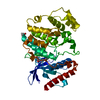
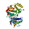
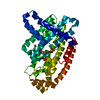
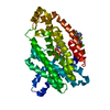
| タイトル | MITOGEN ACTIVATED PROTEIN KINASE 7 IN COMPLEX WITH INHIBITOR |
|---|
 要素 要素 | Mitogen-activated protein kinase 7 |
|---|
 キーワード キーワード | TRANSFERASE / KINASE / INHIBITOR |
|---|
| 機能・相同性 |  機能・相同性情報 機能・相同性情報
Signalling to ERK5 / negative regulation of response to cytokine stimulus / negative regulation of heterotypic cell-cell adhesion / calcineurin-NFAT signaling cascade / cellular response to laminar fluid shear stress / Gastrin-CREB signalling pathway via PKC and MAPK / ERKs are inactivated / mitogen-activated protein kinase binding / negative regulation of calcineurin-NFAT signaling cascade / ERK/MAPK targets ...Signalling to ERK5 / negative regulation of response to cytokine stimulus / negative regulation of heterotypic cell-cell adhesion / calcineurin-NFAT signaling cascade / cellular response to laminar fluid shear stress / Gastrin-CREB signalling pathway via PKC and MAPK / ERKs are inactivated / mitogen-activated protein kinase binding / negative regulation of calcineurin-NFAT signaling cascade / ERK/MAPK targets / negative regulation of smooth muscle cell apoptotic process / positive regulation of protein metabolic process / RET signaling / MAP kinase activity / mitogen-activated protein kinase / regulation of angiogenesis / negative regulation of extrinsic apoptotic signaling pathway in absence of ligand / cellular response to transforming growth factor beta stimulus / negative regulation of endothelial cell apoptotic process / negative regulation of oxidative stress-induced intrinsic apoptotic signaling pathway / enzyme inhibitor activity / PML body / cellular response to growth factor stimulus / negative regulation of inflammatory response / cellular response to hydrogen peroxide / MAPK cascade / adenylate cyclase-activating G protein-coupled receptor signaling pathway / Senescence-Associated Secretory Phenotype (SASP) / cell differentiation / intracellular signal transduction / protein serine kinase activity / protein serine/threonine kinase activity / signal transduction / positive regulation of transcription by RNA polymerase II / nucleoplasm / ATP binding / nucleus / cytoplasm / cytosol類似検索 - 分子機能 Mitogen-activated protein (MAP) kinase, conserved site / MAP kinase signature. / : / Phosphorylase Kinase; domain 1 / Phosphorylase Kinase; domain 1 / Transferase(Phosphotransferase) domain 1 / Transferase(Phosphotransferase); domain 1 / Serine/threonine-protein kinase, active site / Serine/Threonine protein kinases active-site signature. / Protein kinase domain ...Mitogen-activated protein (MAP) kinase, conserved site / MAP kinase signature. / : / Phosphorylase Kinase; domain 1 / Phosphorylase Kinase; domain 1 / Transferase(Phosphotransferase) domain 1 / Transferase(Phosphotransferase); domain 1 / Serine/threonine-protein kinase, active site / Serine/Threonine protein kinases active-site signature. / Protein kinase domain / Serine/Threonine protein kinases, catalytic domain / Protein kinase, ATP binding site / Protein kinases ATP-binding region signature. / Protein kinase domain profile. / Protein kinase domain / Protein kinase-like domain superfamily / 2-Layer Sandwich / Orthogonal Bundle / Mainly Alpha / Alpha Beta類似検索 - ドメイン・相同性 Chem-4R0 / Mitogen-activated protein kinase 7類似検索 - 構成要素 |
|---|
| 生物種 |  Homo sapiens (ヒト) Homo sapiens (ヒト) |
|---|
| 手法 |  X線回折 / シンクロトロン / 分子置換 / 解像度: 2.48 Å X線回折 / シンクロトロン / 分子置換 / 解像度: 2.48 Å |
|---|
 データ登録者 データ登録者 | Tucker, J. / Ogg, D.J. |
|---|
 引用 引用 | ジャーナル: Acta Crystallogr D Struct Biol / 年: 2016
タイトル: Discovery of a novel allosteric inhibitor-binding site in ERK5: comparison with the canonical kinase hinge ATP-binding site.
著者: Chen, H. / Tucker, J. / Wang, X. / Gavine, P.R. / Phillips, C. / Augustin, M.A. / Schreiner, P. / Steinbacher, S. / Preston, M. / Ogg, D. |
|---|
| 履歴 | | 登録 | 2015年5月13日 | 登録サイト: RCSB / 処理サイト: PDBE |
|---|
| 改定 1.0 | 2016年5月4日 | Provider: repository / タイプ: Initial release |
|---|
| 改定 1.1 | 2016年5月11日 | Group: Database references |
|---|
| 改定 1.2 | 2024年5月1日 | Group: Data collection / Database references / Refinement description
カテゴリ: chem_comp_atom / chem_comp_bond ...chem_comp_atom / chem_comp_bond / database_2 / pdbx_initial_refinement_model
Item: _database_2.pdbx_DOI / _database_2.pdbx_database_accession |
|---|
|
|---|
 ムービー
ムービー コントローラー


 データを開く
 基本情報
基本情報 構造の表示
構造の表示 ダウンロードとリンク
 その他のダウンロード


 PDBj
PDBj


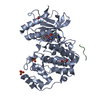
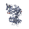
 集合体
集合体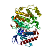

 Spodoptera frugiperda (ツマジロクサヨトウ)
 分子量: 92.094 Da / 分子数: 4 / 由来タイプ: 合成 / 式: C3H8O3
 分子量: 308.767 Da / 分子数: 1 / 由来タイプ: 合成 / 式: C13H17ClN6O
 分子量: 18.015 Da / 分子数: 261 / 由来タイプ: 天然 / 式: H2O
 試料調製
試料調製 / ビームライン: I04 / 波長: 0.979 Å
 解析
解析