 Movie
Movie Controller

[English] 日本語
 Yorodumi
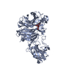
- PDB-4zrs: Crystal structure of a cloned feruloyl esterase from a soil metag... -
+ Open data

- Basic information
| Entry | Database: PDB / ID: 4zrs | |||||||||
|---|---|---|---|---|---|---|---|---|---|---|
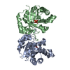
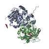
| Title | Crystal structure of a cloned feruloyl esterase from a soil metagenomic library | |||||||||
 Components Components | Esterase | |||||||||
 Keywords Keywords | HYDROLASE / feruloyl esterase / metagenomic library | |||||||||
| Function / homology | : / BD-FAE / Lipase, GDXG, putative histidine active site / Lipolytic enzymes "G-D-X-G" family, putative histidine active site. / : / Alpha/Beta hydrolase fold / hydrolase activity / Esterase Function and homology information Function and homology information | |||||||||
| Biological species |  uncultured bacterium (environmental samples) uncultured bacterium (environmental samples) | |||||||||
| Method |  X-RAY DIFFRACTION / MOLECULAR REPLACEMENT / Resolution: 2 Å X-RAY DIFFRACTION / MOLECULAR REPLACEMENT / Resolution: 2 Å | |||||||||
 Authors Authors | Xie, W. / Chen, R. / Cao, L. / Liu, Y. | |||||||||
| Funding support |  China, 2items China, 2items
| |||||||||
 Citation Citation | Journal: J.Agric.Food Chem. / Year: 2015 Title: Enhancing the Thermostability of Feruloyl Esterase EstF27 by Directed Evolution and the Underlying Structural Basis Authors: Cao, L.C. / Chen, R. / Xie, W. / Liu, Y.H. | |||||||||
| History |
|
- Structure visualization
| Structure viewer | Molecule: MolmilJmol/JSmol |
|---|
- Downloads & links
-Download
| PDBx/mmCIF format | 4zrs.cif.gz | 138.9 KB | Display | PDBx/mmCIF format |
|---|---|---|---|---|
| PDB format | pdb4zrs.ent.gz | 106 KB | Display | PDB format |
| PDBx/mmJSON format | 4zrs.json.gz | Tree view | PDBx/mmJSON format | |
| Others |  Other downloads Other downloads |
-Validation report
| Arichive directory | https://data.pdbj.org/pub/pdb/validation_reports/zr/4zrsftp://data.pdbj.org/pub/pdb/validation_reports/zr/4zrs | HTTPS FTP |
|---|
-Related structure data
| Related structure data |  4nspS S: Starting model for refinement |
|---|---|
| Similar structure data |
-Links
 PDBj
PDBj- Assembly
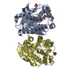
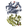
| Deposited unit | 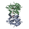
| ||||||||||||
|---|---|---|---|---|---|---|---|---|---|---|---|---|---|
| 1 |
| ||||||||||||
| Unit cell |
| ||||||||||||
| Noncrystallographic symmetry (NCS) | NCS oper:
|
-Components
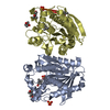
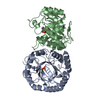
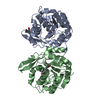
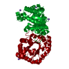
| #1: Protein | Mass: 33453.750 Da / Num. of mol.: 2 / Mutation: T27I, S84L, V161I, H195L, G243C, A259V Source method: isolated from a genetically manipulated source Source: (gene. exp.) uncultured bacterium (environmental samples)Plasmid: pET28a(+) / Production host: #2: Chemical |   Mass: 92.094 Da / Num. of mol.: 2 / Source method: obtained synthetically / Formula: C3H8O3 Mass: 92.094 Da / Num. of mol.: 2 / Source method: obtained synthetically / Formula: C3H8O3#3: Water | ChemComp-HOH / |  Mass: 18.015 Da / Num. of mol.: 615 / Source method: isolated from a natural source / Formula: H2O Mass: 18.015 Da / Num. of mol.: 615 / Source method: isolated from a natural source / Formula: H2O |
|---|
-Experimental details
-Experiment
| Experiment | Method: X-RAY DIFFRACTION |
|---|
- Sample preparation
| Crystal | Density Matthews: 2.71 Å3/Da / Density % sol: 54.6 % |
|---|---|
| Crystal grow | Temperature: 298 K / Method: vapor diffusion, sitting drop / pH: 7.5 / Details: 1.2 M ammonium sulfate, 100 mM HEPES |
-Data collection
| Diffraction | Mean temperature: 100 K |
|---|---|
| Diffraction source | Source: ROTATING ANODE / Type: OTHER / Wavelength: 1.54056 Å |
| Detector | Type: OXFORD ONYX CCD / Detector: CCD / Date: May 9, 2014 |
| Radiation | Protocol: SINGLE WAVELENGTH / Monochromatic (M) / Laue (L): M / Scattering type: x-ray |
| Radiation wavelength | Wavelength: 1.54056 Å / Relative weight: 1 |
| Reflection | Resolution: 2→65.35 Å / Num. obs: 45906 / % possible obs: 99.7 % / Redundancy: 3.3 % / Rmerge(I) obs: 0.104 / Net I/σ(I): 10.6 |
| Reflection shell | Resolution: 2→2.11 Å / Redundancy: 3.2 % / Rmerge(I) obs: 0.435 / Mean I/σ(I) obs: 3 / % possible all: 100 |
- Processing
| Software |
| ||||||||||||||||||||||||||||||||||||||||||||||||||||||||||||||||||||||||||||||||||||||||||||||||||||||||||||||||||||||||||||||||||||||||||||||||||||||||||||||||||||||||||||||||||||||
|---|---|---|---|---|---|---|---|---|---|---|---|---|---|---|---|---|---|---|---|---|---|---|---|---|---|---|---|---|---|---|---|---|---|---|---|---|---|---|---|---|---|---|---|---|---|---|---|---|---|---|---|---|---|---|---|---|---|---|---|---|---|---|---|---|---|---|---|---|---|---|---|---|---|---|---|---|---|---|---|---|---|---|---|---|---|---|---|---|---|---|---|---|---|---|---|---|---|---|---|---|---|---|---|---|---|---|---|---|---|---|---|---|---|---|---|---|---|---|---|---|---|---|---|---|---|---|---|---|---|---|---|---|---|---|---|---|---|---|---|---|---|---|---|---|---|---|---|---|---|---|---|---|---|---|---|---|---|---|---|---|---|---|---|---|---|---|---|---|---|---|---|---|---|---|---|---|---|---|---|---|---|---|---|
| Refinement | Method to determine structure: MOLECULAR REPLACEMENT Starting model: 4NSP Resolution: 2→65.35 Å / Cor.coef. Fo:Fc: 0.963 / Cor.coef. Fo:Fc free: 0.937 / SU B: 3.541 / SU ML: 0.097 / Cross valid method: THROUGHOUT / ESU R: 0.142 / ESU R Free: 0.138 / Stereochemistry target values: MAXIMUM LIKELIHOOD / Details: HYDROGENS HAVE BEEN ADDED IN THE RIDING POSITIONS
| ||||||||||||||||||||||||||||||||||||||||||||||||||||||||||||||||||||||||||||||||||||||||||||||||||||||||||||||||||||||||||||||||||||||||||||||||||||||||||||||||||||||||||||||||||||||
| Solvent computation | Ion probe radii: 0.8 Å / Shrinkage radii: 0.8 Å / VDW probe radii: 1.2 Å / Solvent model: MASK | ||||||||||||||||||||||||||||||||||||||||||||||||||||||||||||||||||||||||||||||||||||||||||||||||||||||||||||||||||||||||||||||||||||||||||||||||||||||||||||||||||||||||||||||||||||||
| Displacement parameters | Biso mean: 18.827 Å2
| ||||||||||||||||||||||||||||||||||||||||||||||||||||||||||||||||||||||||||||||||||||||||||||||||||||||||||||||||||||||||||||||||||||||||||||||||||||||||||||||||||||||||||||||||||||||
| Refinement step | Cycle: LAST / Resolution: 2→65.35 Å
| ||||||||||||||||||||||||||||||||||||||||||||||||||||||||||||||||||||||||||||||||||||||||||||||||||||||||||||||||||||||||||||||||||||||||||||||||||||||||||||||||||||||||||||||||||||||
| Refine LS restraints |
|